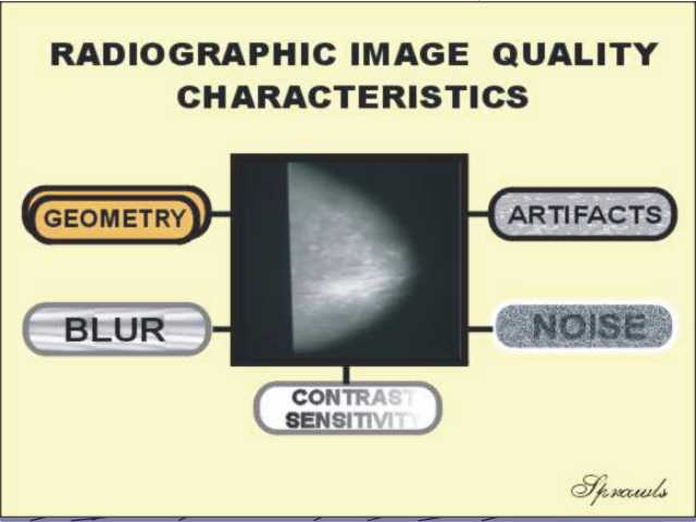
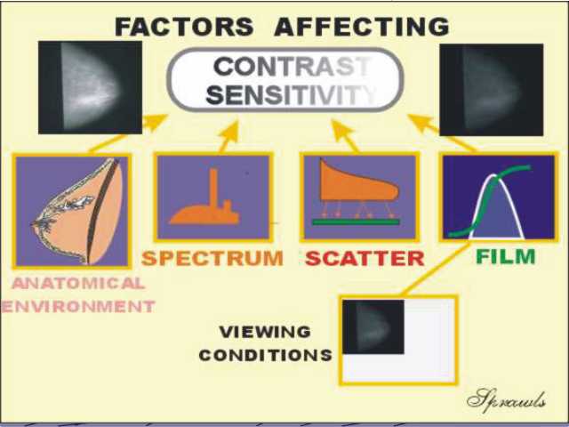
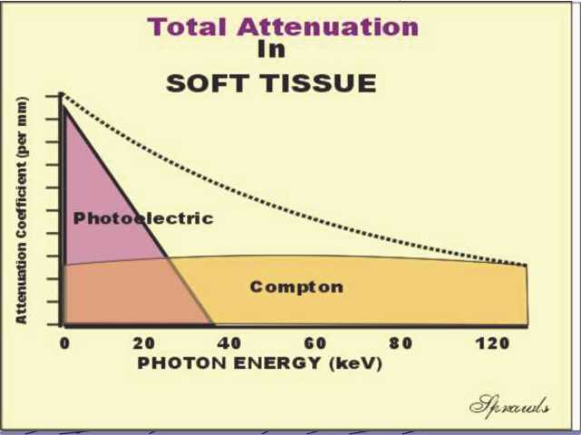
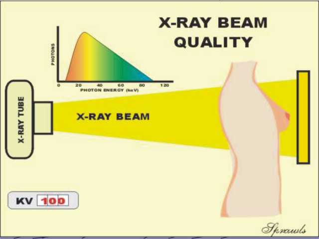
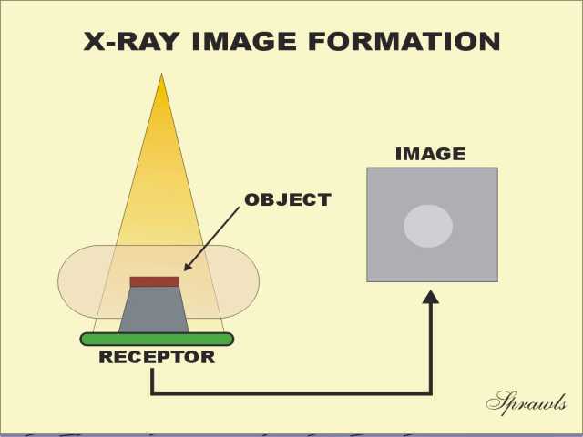
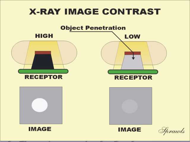
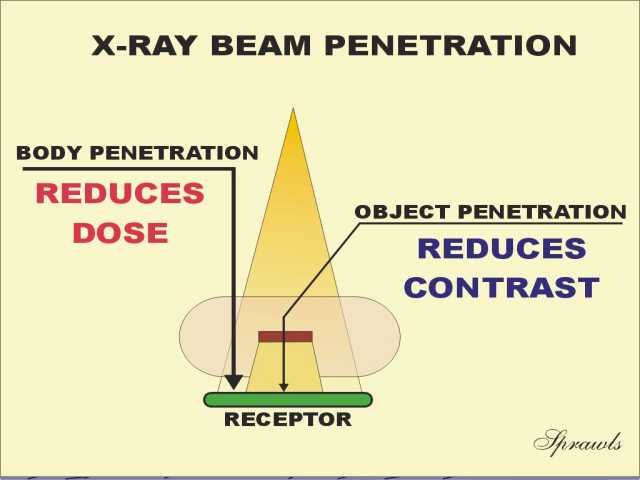
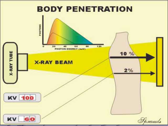
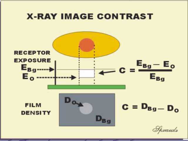
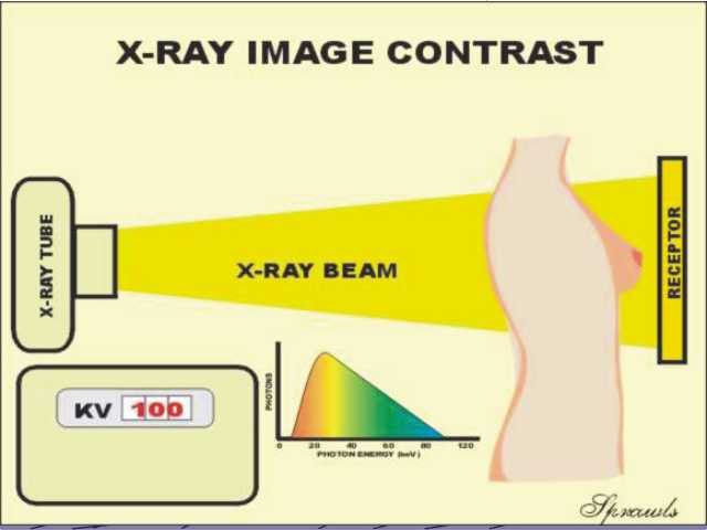
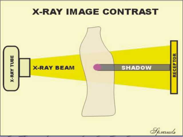
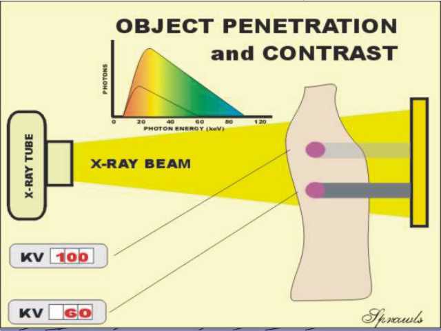
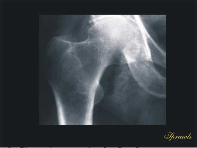
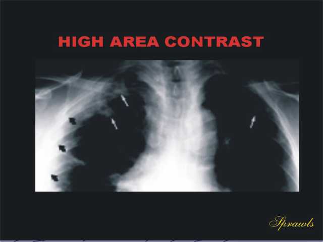
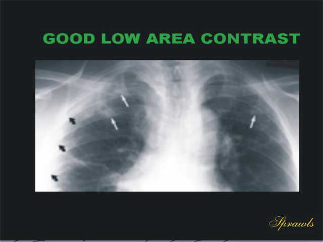
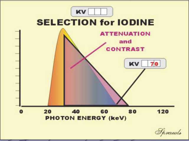
X-ray Image Formation And Contrast

To begin online module, CLICK HERE.


1


BACK

NEXT

The five radiographic image quality characteristics.  Contrast sensitivity is a major characteristic that determines what tissues and body structures are visible in a radiograph.


2


BACK

NEXT

The many factors that have an effect on radiographic contrast sensitivity.


3


BACK

NEXT

The total attenuation by soft tissue is determined by both photoelectric and Compton interactions.  Attenuation generally decreases with increased photon energy because of the energy dependence of the photoelectric effect.


4


BACK

NEXT

The photon energy spectrum of an x-ray beam determines its quality.


5


BACK

NEXT

An x-ray image is formed by the different attenuations of the x-ray beam within a patient's body.  Objects with increased attenuation produce shadows.


6


BACK

NEXT

The image contrast produced by an object depends on its attenuation of the x-ray beam.


7


BACK

NEXT

Generally increased penetration through an object decreases contrast.  In creased penetration through the total body generally decreases the radiation dose to the patient.


8


BACK

NEXT

Penetration through the body is generally increased by increasing the KV.


9


BACK

NEXT

Calculation of the amount of contrast coming from a patient's body and also recorded on film.


10


BACK

NEXT

Changing KV is the principle control of contrast in radiography.


11


BACK

NEXT

The x-ray image of an object in a patient's body is in the form of a shadow.


12


BACK

NEXT

Object penetration and contrast can be changed by changing KV.


13


BACK

NEXT

The x-ray attenuation and total body penetration changes with photon energy.


14


BACK

NEXT

An object must have some form of physical contrast in order to be visible in an x-ray image.


15


BACK

NEXT

The forms of physical contrast that contribute to contrast in x-ray imaging.


16


BACK

NEXT

The penetration through soft tissue can be changed by changing the KV which changes the spectrum.

.


17


BACK

NEXT

X-ray spectra produced by different KV values.


18


BACK

NEXT

The spectrum of an x-ray beam is determined by: the anode material, the KV value, and the filtration.


19


BACK

NEXT

The chest has high physical contrast primarily because of the air within the lungs.


20


BACK

NEXT

Good chest radiographs are produced by using a high KV value and a heavily filtered beam with a spectrum that gives good penetration.


21


BACK

NEXT

An image showing good penetration through the bones.


22


BACK

NEXT

An image with high area contrast is not desirable.


23


BACK

NEXT

Generally, relatively low contrast between large areas within the body produce better local object contrast and visibility.


24


BACK

NEXT

Bone image.


25


BACK

NEXT

Good contrast of iodine requires a spectrum with a significant amount of radiation above the K-edge energy of iodine.  This is usually produced with KV values in the range of 65-75.


26


BACK

NEXT

KV values for the different radiographic procedures.


27


BACK

NEXT

The breast generally has low physical contrast.  It is in the form of relatively small differences in density among the soft tissues and the low contrast of small calcifications.


28


BACK

NEXT

The x-ray spectrum used for mammography is determined by the anode material, selected KV value, and type of filter.


29


BACK

NEXT

The spectrum and the resulting breast penetration can be changed by alternating between different filter materials.


30


BACK

NEXT


31

The End

BACK


To return to the beginning,
CLICK HERE