|
|
Online Module |
|
|
Online Module |
Medical Image Characteristics and Quality Factors
To begin module, CLICK HERE.
To go to a specific topic, click on topic title below.
|
It is formed by the imaging modalities that use various forms of radiation and energy to open the body to visualization. |
|
|
|
Generally, the factors that affect image quality for any specific imaging method have a direct effect on one or more of these specific characteristics. A knowledge of these relationships is necessary for:
|
|
For medical imaging we have a choice of:
The general principles and the technology of each of
these methods determine the image characteristics. |
|
For each imaging method there are certain technique or protocol factors that must be adjusted by the operator before performing the imaging procedure. It is the selected values of these factors that determine the specific image characteristics. For most imaging methods there is a range of image quality within which the equipment can be operated. The actual quality within this range is determined by how the equipment is operated. That raises an interesting question.... If the image quality can be adjusted, why not always adjust for maximum quality and visibility? Answer.... In medical imaging there are many trade-offs and compromises that must be considered.
That is why the technique factors and imaging protocol for each image acquisition should be selected to produce an optimized procedure, based on the specific clinical requirements for visibility.
An optimized protocol
produces a balance among the image characteristics (for example,
blurring and noise) and uses the radiation dose that is necessary to
produce the required image quality. |
|
Just as we use the microscope to view small things, and the telescope and television to view things at a distance, medical imaging equipment extends our vision into the normally invisible regions of the human body. This is a useful concept because we can use some of the same characteristics used to evaluate human vision to evaluate a medical imaging procedure. Now, Let's test our vision.... |
|
Can you see the G, the H, the I? Make a note of your test results. Before we move on, let's notice the characteristic that is changing as we go up the alphabet. It is the CONTRAST of the letters. The letter A has a very high contrast in relationship to its background, and is very visible and easy to see. As we read up the alphabet, it is the contrast of the letters that is decreasing, and reducing their visibility. What characteristic of our vision have we just tested? |
|
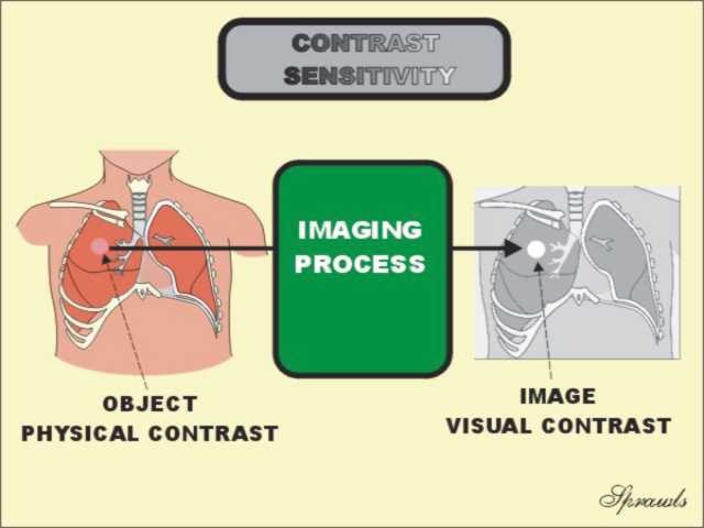
We have just tested the CONTRAST SENSITIVITY of our visual system. Contrast sensitivity is the characteristic of an imaging system that determines the lowest contrast object that can be visualized. It is one of the most significant characteristics of all medical imaging methods. In medical imaging it is often necessary to visualize objects and structures in the body that themselves have very low physical contrast. Soft tissue masses surrounded by normal soft tissue are good examples.
|
|
The relationship of the visual contrast to the physical contrast is determined by the contrast sensitivity of the process. Contrast sensitivity is generally expressed as the lowest physical contrast that produces the necessary image contrast for visibility. Contrast resolution is an alternate term that is sometimes used for this general characteristic of a medical imaging process. |
|
When the contrast sensitivity is low the result will be low image contrast and low visibility. We cannot always detect low contrast sensitivity by just looking at an image. We might have an image that appears to have high visual contrast. Such an image might be showing some of the high contrast objects like, like bones and bullets, but not showing low contrast objects like soft tissue masses. |
|
Each imaging modality has contrast sensitivity characteristics that relate to the type of physical contrast it can image and the lowest physical contrast that can be visualized. The contrast sensitivity that is actually achieved then depends on the operating factors used for a specific procedure. |
|
Here we can compare two imaging procedures, one with a low and the other with a higher contrast sensitivity. As shown on the left, objects in the body have a wide range of physical contrast. Bones, barium, and bullets (the three "B"s) have very high physical contrast compared to surrounding soft tissues. Most soft tissue objects, such as cancers, usually have a very low physical difference (contrast) relative to the surrounding tissue. When imaged with a low contrast sensitivity, only the high contrast objects are visible. High contrast sensitivity is required to visualize the many low contrast structures, objects, and signs of pathology in the body. |
|
Very low image contrast, as shown on the left, usually results in low contrast sensitivity and low visibility. In most cases, there is usually an optimum image contrast that gives the best visibility of objects throughout the image. Optimum contrast is obtained by the right combination of film type, film processing, and level of exposure. In digital images, optimum contrast is obtained with the appropriate digital processing and adjustment of display factors, especially the windowing. |
|
The contrast of an object is expressed relative to its surrounding background. That is what determines its visibility. For an image recorded on film, the contrast of a specific object is the difference in the film density (darkness) values. These can be measured with a densitometer. Note: Measuring object contrast in a test film with a densitometer is a routine function in mammography quality assurance procedures. For displayed images the contrast of a specific object is the ratio of the brightness of the object to the brightness of the background. |
|
Physical contrast is the difference in some physical characteristic of the tissues or other materials in the body. The physical characteristics that produce visible object contrast is different for the different imaging modalities. For x-ray imaging, including CT, the two sources of physical contrast are:
Radiographs and CT images are generally images
showing differences in density within the body. CT images also display differences in density. However, CT has a very high contrast sensitivity compared to radiography and can display relatively small differences in tissue density, as in the head. When there is not a sufficient difference in density within the body to produce visible contrast (like between fluids and soft tissue) compounds of either iodine or barium can be administered to enhance contrast. Iodine and barium are good x-ray absorbers, not so much because of their density, but because of their atomic numbers (atomic size). As described in later modules the arrangement of the electrons (their binding energies) in these two elements makes them especially good absorbers and sources of contrast. |
|
A radiographic technique that has low contrast sensitivity is used for chest imaging because of the high level of physical contrast (variation in density) that is present. Mammography requires a technique with high contrast sensitivity because of the very low physical contrast in the breast. There are several factors that determine the contrast sensitivity of a radiographic/mammographic procedure. The major factor is the selected KV value as described in later modules. |
|
Different modes are used to image the two different types of physical contrast. The B (Brightness) mode displays the intensity of reflections or echoes as image brightness. A bright spot indicates a structure surface or boundary that is producing strong echoes (because of a large difference in acoustic impedance). The Doppler mode displays flowing blood. |
|
Physical contrast depends on the differential uptake and distributions associated with the various physiological functions and pathological conditions. The imaging equipment must be adjusted to maximize the contrast sensitivity to the specific radionuclide that is being used.
The critical adjustment is that of the energy window in
the Pulse Height Analyzer (PHA) of the gamma camera. |
|
Most imaging is based on the three magnetic (physical) characteristics of tissue:
The contrast sensitivity to a specific characteristic is set by the selected protocol factor values. Three forms of fluid movement are other physical characteristics that can be sources of contrast in MRI. The visualization of each requires special imaging techniques. |
|
Would you agree that the image on the left is more blurred? Now let's consider the effect of this blurring on visibility. In the image on the right, with much less blurring, we have much better visibility of the anatomical details. It is the blurring in medical images that limits visibility of detail. There is some amount of blurring in all medical images. A knowledge of this blurring effect and how to control it is necessary for optimizing imaging procedures to insure appropriate visibility. |
|
The letters and numbers become smaller as we move down the chart, this represents increasing detail. Most of us with normal or corrected vision can read all rows. What would we see (or not see) if we had very blurred vision? |
|
This image has been blurred to demonstrate a very important concept. Blurring reduces the visibility of small objects and detail. This applies to all forms of medical imaging. |
|
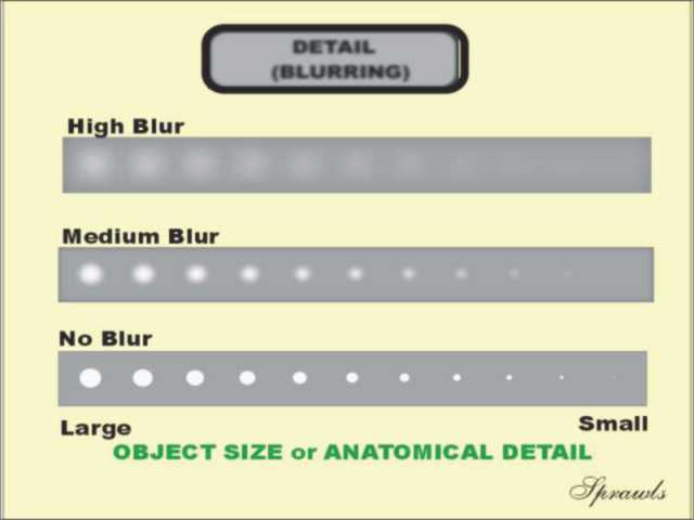
The smaller objects represent increased anatomical detail. Here we have arranged a series of objects by size or detail. We will now use this to observe the effects of blurring. In this image with relatively little blur, we can see all of the objects. Now, let's blur the image. |
|
Notice that even in the blurred images, the large objects are visible. Blurring places a limit on the smallest objects that can be seen. Blurring limits visibility of detail. |
|
|
|
The amount of blurring (and visibility of detail) in a specific imaging procedure is determined by a combination of two factors:
|
|
The difference in the amount of blurring from one modality to another is because of the different principles and methods of image formation and the design characteristics of the equipment. The scale shows the size of the blur and the associated maximum spatial resolution which relates to visibility of detail. Notice that of all of the modalities, radiography produces images with the least blurring and therefore the best visibility of detail. Mammography is the special radiographic procedure that has the least blurring (approximately 0.12 mm) and is capable of producing images of calcifications in the range of 0.1-0.2 mm). |
|
There are three specific effects of blurring in medical imaging:
The amount (size) of blurring in a specific imaging procedure is determined by:
In later modules we will see that blurs has different shapes, depending on the source of the blurring. |
|
All medical images have some amount of
visual noise. The image on the left has a relatively low amount of noise and is the range that would be acceptable for a clinical study. The image on the right has a high level of noise and would not be acceptable for a clinical study. |
|
There are different levels (low, medium, and high) of noise in the three columns. Notice that the effect of the noise is to reduce the visibility of the low contrast objects. |
|
This illustration is a Contrast-Detail Diagram. Notice that the objects are arranged according to decreasing size (detail) from left to right, and according to decreased contrast from bottom to top. The larger and high contrast objects in the lower left region should be visible under most imaging conditions. Think of noise and blur as two characteristics that together produce a "curtain of invisibility". Noise reduces visibility of low contrast objects. Blur reduces visibility of small objects. Typically, most small anatomical objects also have relatively low contrast and their visibility is reduced by both noise and blurring. |
|
Noise is reduced by imaging with a greater concentration of photons which generally requires a higher radiation dose to the patient. The noise level is also affected by the selected values of some of the imaging protocol factors as illustrated here for CT. Adjusting an imaging procedure to reduce noise usually results in an increased exposure to the patient or an adverse effect on one of the other image characteristics, such as blurring. The factors affecting noise will be described in other modules. |
|
Each view has both advantages and limitations. |
|
Typically an anatomical region is divided into slices. The slices are divided into a matrix (array) of tissue voxels (volume elements). The digital image of the slice is formed as a matrix of pixels (picture elements). The brightness or color displayed in each pixel represents some physical characteristic of the tissue in the voxel.
|
|
Characteristics of anatomical structures and objects that can be distorted include:
|
|
|
|
|
|
A long (72 in) FRD is typically used for chest imaging to minimize magnification and the related distortion. |
|
Each characteristic can have an effect on the visibility of structures and objects within the body. |
|
This is the end of this module. To return to the beginning, CLICK HERE.
|

 The
medical image is a window into the human body.
The
medical image is a window into the human body.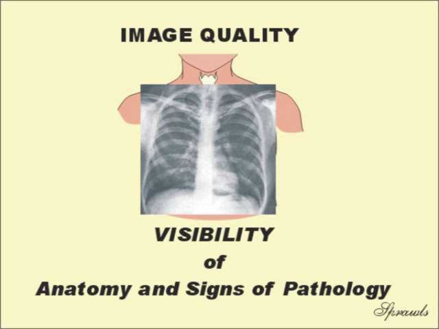 Image
quality is the overall characteristic of a medical image that
determines which objects and structures within a body are visible.
Image
quality is the overall characteristic of a medical image that
determines which objects and structures within a body are visible.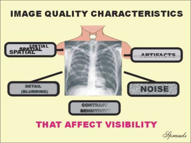 Image
quality, and visibility of the internal body, is determined by a
combination of five more specific image characteristics. They are:
Image
quality, and visibility of the internal body, is determined by a
combination of five more specific image characteristics. They are: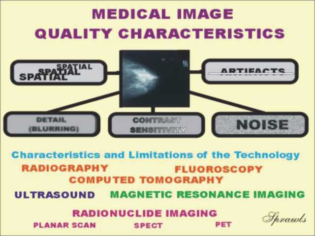 The
first thing that determines the characteristics and quality of a medical
image is the imaging method or technology used to produce the image.
The
first thing that determines the characteristics and quality of a medical
image is the imaging method or technology used to produce the image.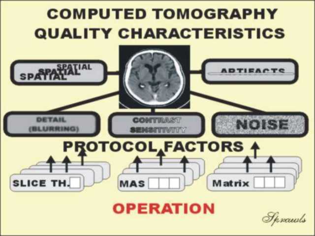 For
each imaging method (CT is illustrated here) the specific image quality
characteristics are determined by how the equipment is operated.
For
each imaging method (CT is illustrated here) the specific image quality
characteristics are determined by how the equipment is operated.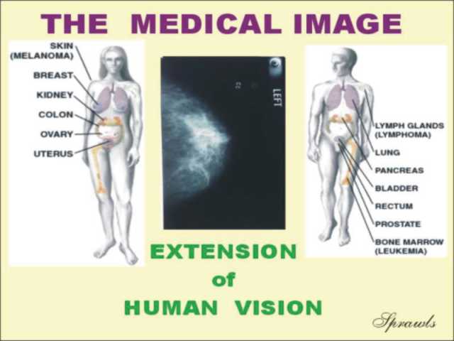 Medical
imaging technology is actually an extension of our human vision.
Medical
imaging technology is actually an extension of our human vision.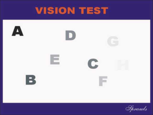 The
test is to look at the chart and read as far up the alphabet that you
can.
The
test is to look at the chart and read as far up the alphabet that you
can.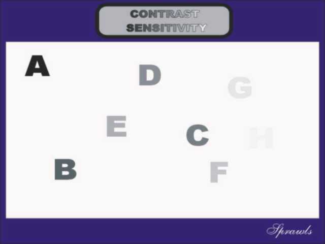
 The
function of all medical imaging procedures is to:
The
function of all medical imaging procedures is to: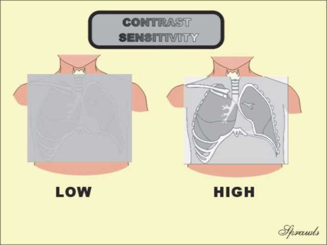 Visibility
of anatomy and signs of pathology depend on the contrast that is
produced and displayed in the image.
Visibility
of anatomy and signs of pathology depend on the contrast that is
produced and displayed in the image.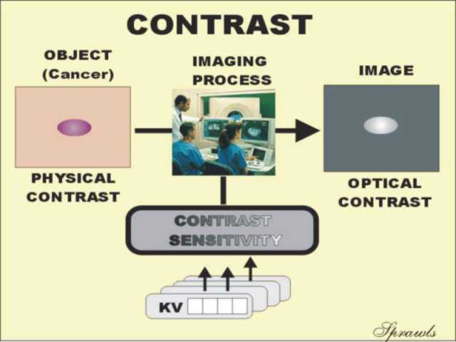 Contrast
sensitivity, that is the ability to convert and transfer contrast, is
determined by two factors:
Contrast
sensitivity, that is the ability to convert and transfer contrast, is
determined by two factors: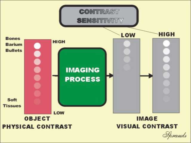
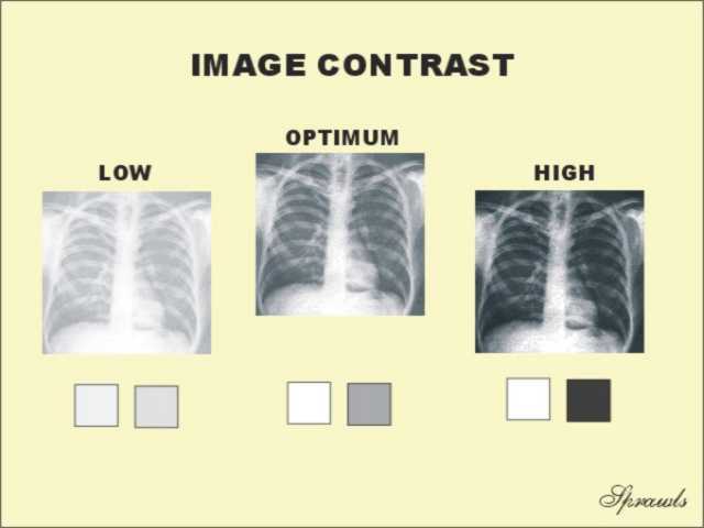 The
objective is not to always have the highest possible contrast in an
image. If there is high contrast between areas in an image (area
contrast) visibility might be reduced in the very light and dark areas.
This is especially true for images recorded on film because the contrast
sensitivity of film is reduced in the light (low exposure) and dark
(high exposure) areas.
The
objective is not to always have the highest possible contrast in an
image. If there is high contrast between areas in an image (area
contrast) visibility might be reduced in the very light and dark areas.
This is especially true for images recorded on film because the contrast
sensitivity of film is reduced in the light (low exposure) and dark
(high exposure) areas.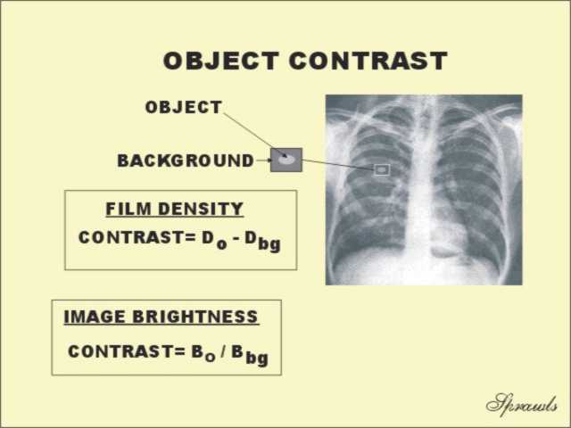 The
contrast of individual objects within an image can be measured and given
a numerical value.
The
contrast of individual objects within an image can be measured and given
a numerical value.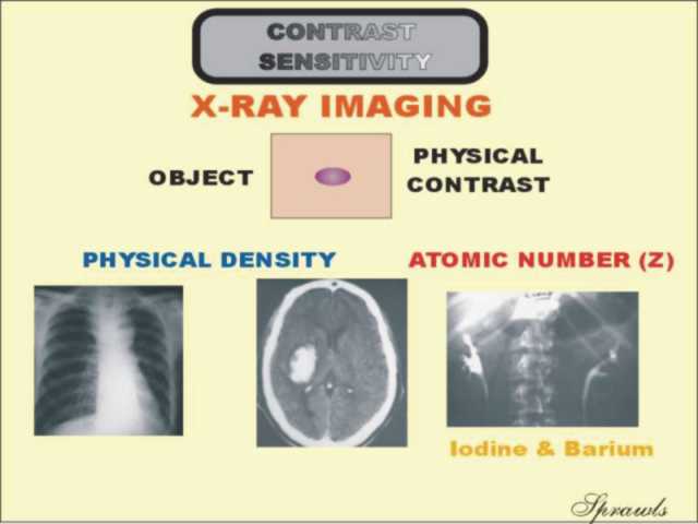 Objects
and structures in the body must have some form of physical contrast in
order to produce visible contrast in an image.
Objects
and structures in the body must have some form of physical contrast in
order to produce visible contrast in an image.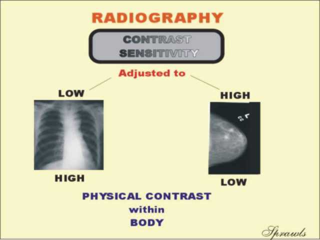 The
contrast sensitivity of an imaging
procedure must be adjusted according to the the amount of
physical contrast in a specific anatomical
region.
The
contrast sensitivity of an imaging
procedure must be adjusted according to the the amount of
physical contrast in a specific anatomical
region.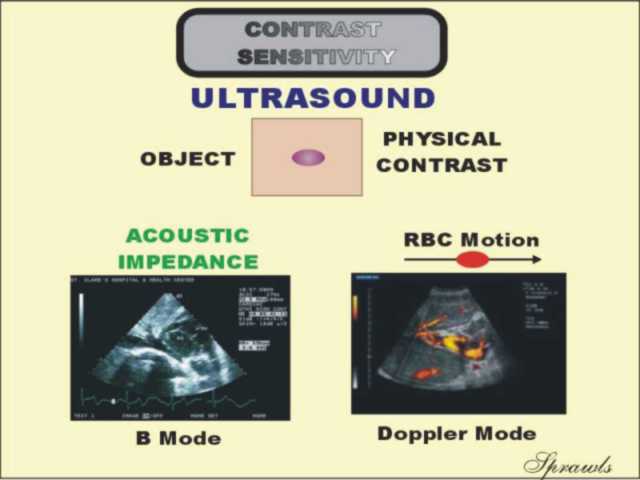 In
ultrasound imaging there are two principle source of physical contrast
that can be imaged:
In
ultrasound imaging there are two principle source of physical contrast
that can be imaged: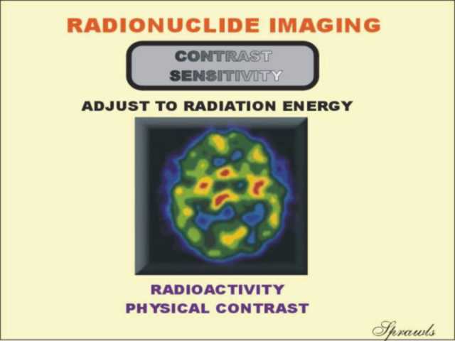 Physical
contrast in all types of radionuclide imaging (including SPECT and PET)
is the variation of radioactivity throughout a body section.
Physical
contrast in all types of radionuclide imaging (including SPECT and PET)
is the variation of radioactivity throughout a body section.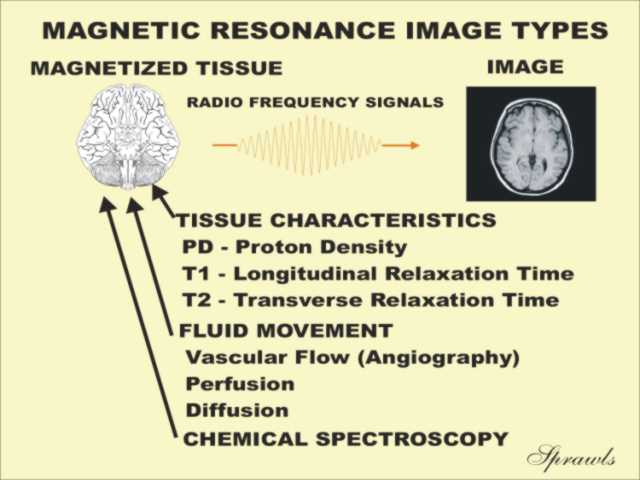 MRI
can produce visible images of several different physical characteristics
as illustrated here.
MRI
can produce visible images of several different physical characteristics
as illustrated here.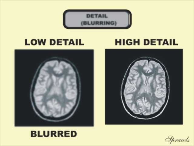 Compare
the two images shown here. What difference do you see?
Compare
the two images shown here. What difference do you see?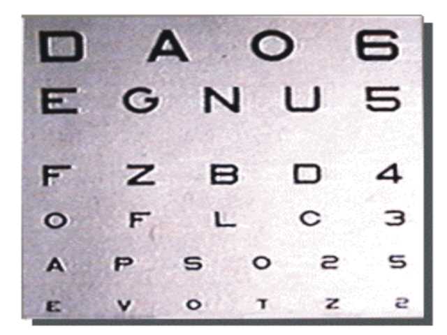 This
test chart can be used to evaluate visibility of detail.
This
test chart can be used to evaluate visibility of detail.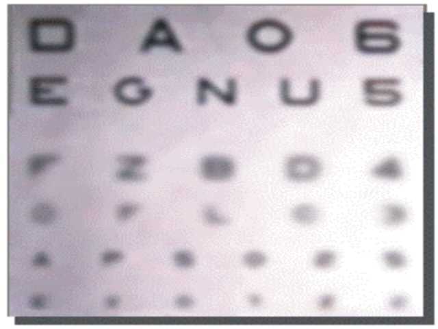 It
is not your vision that is the problem!
It
is not your vision that is the problem!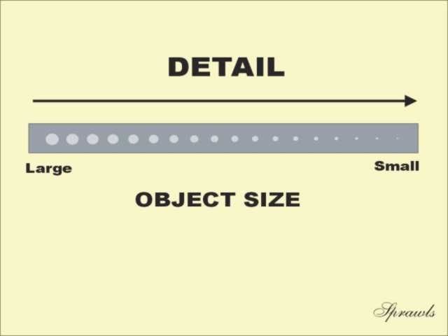 Objects
in the body cover a wide range of sizes.
Objects
in the body cover a wide range of sizes.
 These
images show the effect of blurring.
These
images show the effect of blurring.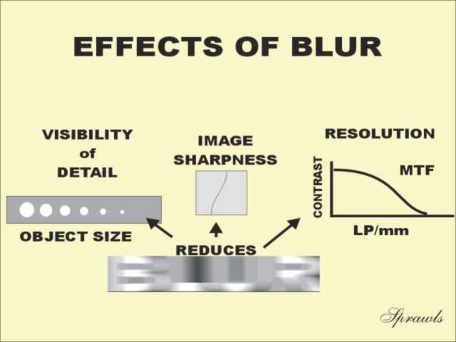 There
are three specific effects of blurring.
There
are three specific effects of blurring.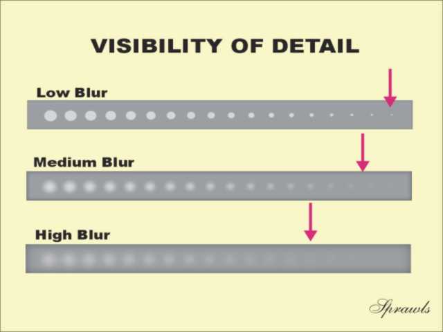 The
clinical significance of the blurring that occurs in all medical imaging
procedures is that it places a limit on the smallest object (anatomical
detail, sign, etc) that will be visible.
The
clinical significance of the blurring that occurs in all medical imaging
procedures is that it places a limit on the smallest object (anatomical
detail, sign, etc) that will be visible.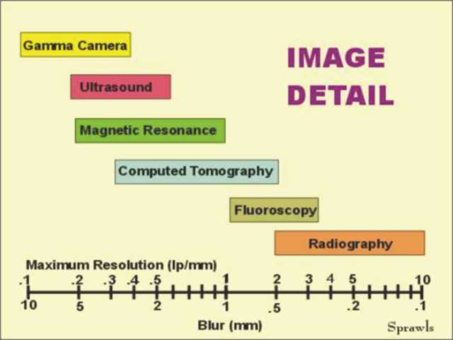 There
is a range of blur values for each imaging modality as illustrated here.
Within that range, the blurring in a specific imaging procedure is
determined by the selection of technique and protocol factors.
There
is a range of blur values for each imaging modality as illustrated here.
Within that range, the blurring in a specific imaging procedure is
determined by the selection of technique and protocol factors.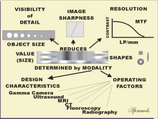 Summary
 Compare
the two images, what difference do you see?
Compare
the two images, what difference do you see?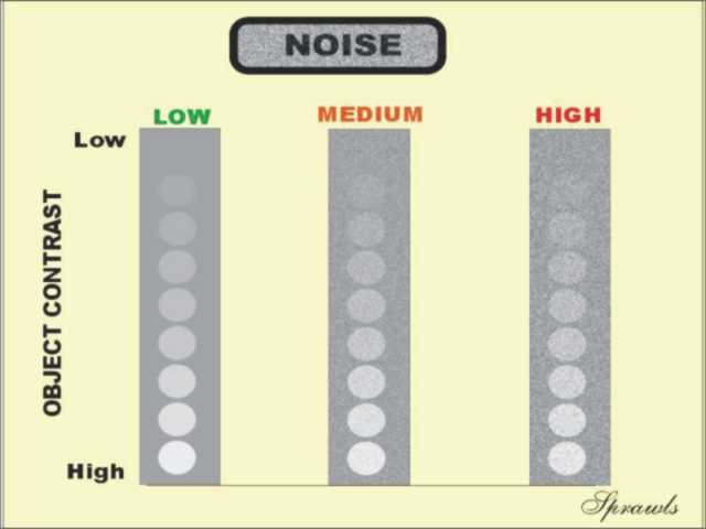 Each
column contains a series of objects ranging from high contrast (bottom)
to very low contrast (top).
Each
column contains a series of objects ranging from high contrast (bottom)
to very low contrast (top).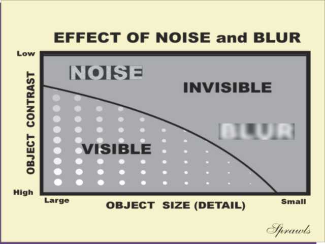 Both
blur and noise
are generally undesirable image characteristics that reduce visibility
of specific objects.
Both
blur and noise
are generally undesirable image characteristics that reduce visibility
of specific objects.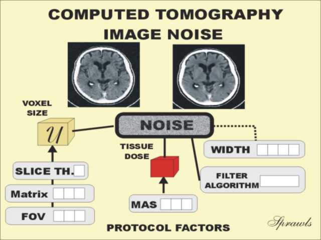 The
most significant source of noise in x-ray imaging (including CT) and
radionuclide imaging (including SPECT and PET) is the random statistical
nature of the photons.
The
most significant source of noise in x-ray imaging (including CT) and
radionuclide imaging (including SPECT and PET) is the random statistical
nature of the photons.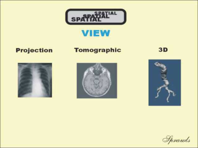 Most
medical images provide one of three possible views of the human body:
Most
medical images provide one of three possible views of the human body: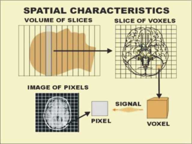 Most
of the tomographic imaging methods, CT, SPECT, PET, and MRI (illustrated
here) have some common spatial characteristics.
Most
of the tomographic imaging methods, CT, SPECT, PET, and MRI (illustrated
here) have some common spatial characteristics.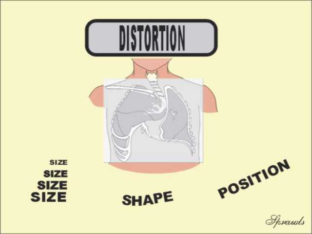 Images
do not always display the true spatial or geometric characteristics of a
body section.
Images
do not always display the true spatial or geometric characteristics of a
body section.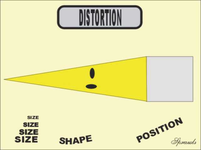 In
radiography most distortion comes from the variation in magnification
with the different object locations in the body and the orientation of
objects with respect to the direction of the x-ray beam.
In
radiography most distortion comes from the variation in magnification
with the different object locations in the body and the orientation of
objects with respect to the direction of the x-ray beam.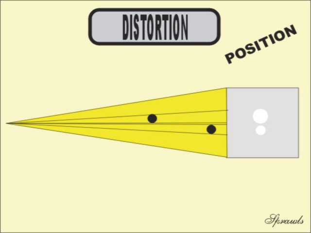 The
relative size and position of objects is distorted by the
projection imaging method as used in
radiography and fluoroscopy.
The
relative size and position of objects is distorted by the
projection imaging method as used in
radiography and fluoroscopy.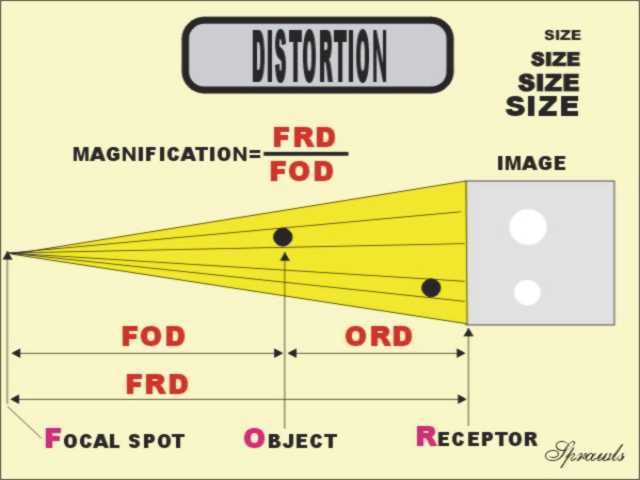 The
magnification of an object is determined by the ratio of distances as
shown.
The
magnification of an object is determined by the ratio of distances as
shown.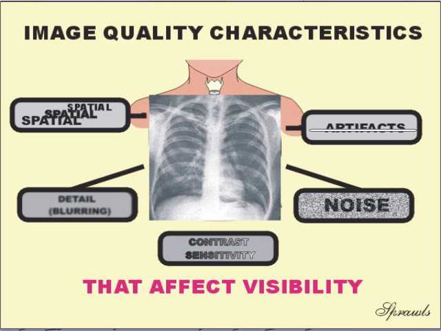 The
general quality of an image is determined
by the combination of the five fundamental characteristics.
The
general quality of an image is determined
by the combination of the five fundamental characteristics.