-
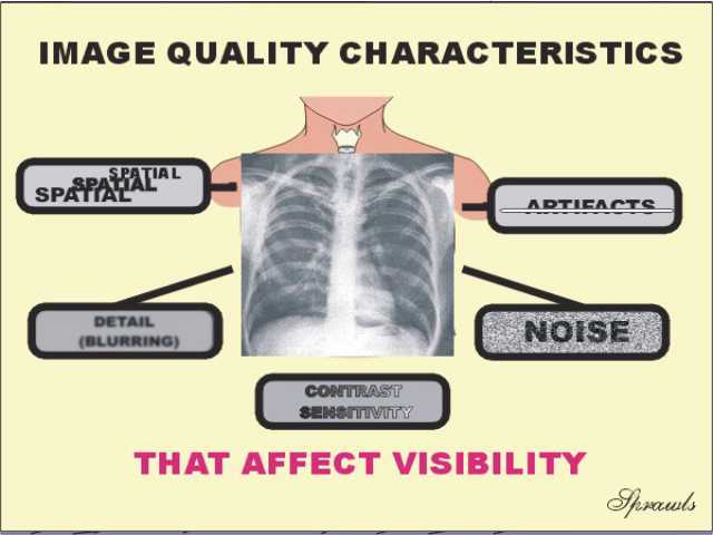
Identify the five basic characteristics of an image that
determine its quality.
-
Describe generally how each characteristic affects
visibility of anatomical structures and signs of pathology.
-
While looking at a chest radiograph, explain the concept of
object and area contrast.
-
While looking at a chest radiograph, identify areas in which
object contrast appears to be reduced because the areas are either too light or
too dark.
-
Explain the concept of contrast sensitivity and it's
significance in medical imaging.
-
Compare the contrast sensitivity characteristics of
radiography and MRI for visualization of soft tissue.
-
Identify the radiographic procedure that requires the
highest contrast sensitivity and explain why.
-
While looking at radiographs, identify some of the objects
that represent anatomical detail, estimate their sizes.
-
Describe the basic relationship between image blurring and
visibility of detail.
-
Compare and rank the various medical imaging modalities with
respect to blurring and visibility of detail. NOTE: In later
modules you will learn the factors in each imaging modality that have an effect
on blurring and visibility of detail.
-
Describe the appearance of image noise.
-
While looking at images from each modality identify the image
noise, Identify the modalities that appear to produce images with relatively low
noise, with relatively high noise.
-
Describe the effect of noise on visibility of objects within
an image and identify the object characteristic that makes it have reduced
visibility in the presence of noise. NOTE: In later modules you
will learn the factors in each imaging modality that produce and have an effect
on the noise.
-
Define and describe image artifact.
-
X Recognize common image artifacts in images from each
modality. NOTE: In later modules you will learn the factors in each
imaging modality that produce and have an effect on artifacts
-
Identify and describe three possible types of medical image
distortion.
-
Identify and describe three major viewing condition factors
that have an effect on the visibility of objects within a medical image.
-
Describe the factors that produce optimum viewing conditions
for medical images.
-
Describe the special viewing conditions required in
mammography and explain how they are achieved.
-
Explain and compare the two characteristics of a diagnostic
procedure: sensitivity and specificity.
-
Define and briefly describe the clinical significance of:
True Positive, False Positive, True Negative, False Negative.
-
Sketch a receiver operating characteristic (ROC) curve, label
the scales, and describe factors that determine the position of the human
operating point.
-
Interpret the general results of a receiver operating
characteristic (ROC) analysis.
-
Briefly describe the clinical application of ROC analysis.