Patient Exposure and Dose Management
Perry Sprawls, Ph.D.

Online Textbook

Table of Contents


INTRODUCTION AND OVERVIEW
X-RAY EXPOSURE PATTERNS
    Radiography
    Fluoroscopy
    Computed Tomography
RADIATION AND IMAGE QUALITY
FACTORS AFFECTING EXPOSURE AND DOSE
    Receptor Sensitivity
      Intensifying Screens
      Films
       Digital Radiographic Receptors
    Grid
    Tabletop
    Distance
    Tissue Penetration
    Exposure Values
    Beam Limiting
EXPOSURE AND DOSE DETERMINATION
RADIONUCLIDE DOSIMETRY
    Total Energy (Integral Dose)
    Cumulated Activity
    Transition Energy
ABSORBED DOSE
    Electron Radiation
    Photon Radiation
ESTIMATION OF DOSE VALUES
CONCLUSION


INTRODUCTION AND OVERVIEW

CONTENTS

   All medical imaging methods deposit some form of energy in the patient's body. Although the quantity of energy is relatively low, it is a factor that should be given attention when conducting diagnostic examinations. In general, there is more concern for the energy deposited by the ionizing radiations, x-ray and gamma, than for ultrasound energy or radio frequency (RF) energy deposited in magnetic resonance imaging (MRI) examinations. Therefore, this chapter gives major emphasis to the issues relating to the exposure of patients to ionizing radiation.

   Patients undergoing either x-ray or radionuclide examinations are subject to a wide range of exposure levels. One of our objectives is to explore the factors that affect patient exposure. This is followed by an explanation of methods that can be used to determine patient exposure values in the clinical setting.

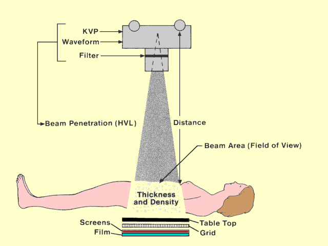
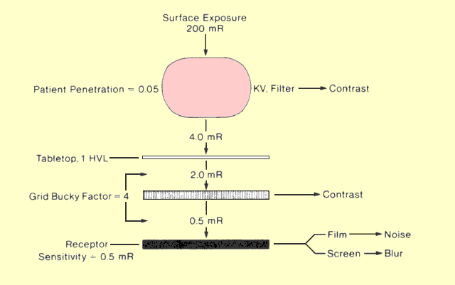
   The following figure identifies the major factors that affect patient exposure during a radiographic procedure. Some factors, such as thickness and density, are determined by the patient. Most of the others are determined by the medical staff. Many of the factors that affect patient exposure also affect image quality. In most instances when exposure can be decreased by changing a specific factor, image quality is also decreased. Therefore, the objective in setting up most x-ray procedures is to select factors that provide an appropriate compromise between patient exposure and image quality.

Factors That Affect Patient Exposure in a Radiographic Procedure

Factors That Affect Patient Exposure in a Radiographic Procedure

      X-RAY EXPOSURE PATTERNS

CONTENTS

   In any x-ray examination, there is considerable variation in exposure from point to point within the patient's body. This must be considered when expressing values for patient exposure. In fact, when exposure values are given, the specific anatomical location of the value should also be stated. Some exposure patterns are characteristic of the different x-ray imaging methods. A review of these patterns will give us some background for considering factors that affect exposure and applying methods to determine actual exposure values.


      Radiography

CONTENTS

   In the typical radiographic examination, the x-ray beam is projected through the patient's body, as shown in the previous figure. The point that receives maximum exposure is the entrance surface near the center of the beam. There are two reasons for this. The primary x-ray beam has not been attenuated by the tissue at this point, and the area is exposed by some of the scattered radiation from the body. The amount of surface exposure produced by the backscatter depends on the spectrum of the primary beam and the size of the exposed area. For typical radiographic situations, scattered radiation can add at least 20% to the surface exposure produced by the primary beam.

   As the x-ray beam progresses through the body, it undergoes attenuation. The rate of attenuation (or penetration) is determined by the photon-energy spectrum (KV and filtration) and the type of tissue (fat, muscle, bone) through which the beam passes. For the purpose of this discussion, we assume a body consisting of homogeneous muscle tissue. In the following figure, lines are drawn to divide the body into HVLs. The exposure is reduced by a factor of one half each time it passes through 1 HVL. The thickness of 1 HVL depends on the photon-energy spectrum. However, for the immediate discussion, we assume that 1 HVL is equivalent to 4 cm of tissue. A 20-cm thick body section consists of 5 HVLs. Therefore, the exposure decreases by one half as it passes through each 4 cm of tissue. At the exit surface, the exposure is a small fraction of the entrance surface exposure.


Typical Exposure Pattern (Depth Dose Curves) for an X-Ray Beam Passing through a Patient's Body

Typical Exposure Pattern (Depth Dose Curves) for an X-Ray Beam Passing through a Patient's Body


   The exposure to a specific organ or point of interest within the direct x-ray beam depends on its proximity to the entrance surface.

   Tissue located outside the primary beam receives some exposure from the scattered radiation produced within the beam area. The scatter exposure to the surrounding tissue is relatively low in comparison to the exposure levels within the primary beam.


      Fluoroscopy

CONTENTS


   The fluoroscopic beam projected through the body will produce a pattern similar to a radiographic beam if the beam remains fixed in one position. If the beam is moved during the procedure, the radiation will be distributed over a large volume of tissue rather than being concentrated in one area. For a specific exposure time, tissue exposure values (roentgens) are reduced by moving the beam, but the total radiation (R - cm2) into the body is not changed. This was illustrated in the figure titled, "Exposure" (in the section titled, "Radiation Quantities and Units").


   Computed Tomography

CONTENTS


   In computed tomography (CT) two factors are associated with exposure distribution and must be considered: (1) the distribution within an individual slice and (2) the effect of imaging multiple slices.

   The rotation of the x-ray beam around the body produces a much more uniform distribution of radiation exposure than a stationary radiographic beam. A typical CT exposure pattern is shown in the figure below. A relatively uniform distribution throughout the slice is obtained if a 360° scan is performed. However, if other scan angles that are not multiples of 360° are used, the exposure distribution will become less uniform.


Typical Exposure (Dose) Pattern Produced with Computed Tomography

Typical Dose Pattern Produced with Computed Tomography


   When multiple slices are imaged, the dose (grays) does not increase in proportion to the number of slices because the radiation is distributed over a larger volume of tissue.  However, when slices are located close together, radiation from one slice can produce additional exposure in adjacent slices because slice edges are not sharply defined  and because of scattered radiation.


RADIATION AND IMAGE QUALITY

CONTENTS

     One of the major compromises that must be made in imaging procedures using ionizing radiation is between patient exposure and image quality. Within certain limits, increasing image quality requires an increase in patient exposure. It is usually the specific image quality requirements that determine the quantity of radiation that must be used in the imaging process. The three basic image quality factors (contrast sensitivity, detail, and noise) are each related to patient exposure. This holds true for both x-ray and nuclear radiation imaging procedures. The variables of an imaging procedure should be selected to produce adequate image quality with the lowest possible radiation exposure.

   We now consider each factor that affects patient exposure and show how it relates to image quality.


FACTORS AFFECTING EXPOSURE AND DOSE

CONTENTS

   The exposure, or dose, to a specific point within a patient's body is determined by a combination of factors. One of the most significant is whether the point in question is in or out of the primary beam. Points not located in the direct beam can receive exposure from scattered radiation, but this is generally much less than the exposure to points within the beam area. The factors that determine exposure levels to points within the body will be discussed in reference to the situation illustrated in the following figure.


Factors That Determine Exposure Values in Radiography

Factors That Determine Exposure Values in Radiography


   Receptor Sensitivity

CONTENTS

   One of the most significant factors is the amount of radiation that must be delivered to the receptor to form a useful image. This is determined by the sensitivity of the receptor. It was shown in the section titled, "Radiographic Receptors" that there is a rather wide range of sensitivity values encountered in radiography. It is generally desirable to use the most sensitive receptor that will give adequate image quality. The exposure to points within the patient's body will be a multiple of the receptor exposure.
The sensitivity of a radiographic receptor is determined both by characteristics of the intensifying screen (as discussed in the section titled, "Radiographic Receptors") and the film (as discussed in the section titled, "The Photographic Process and Film Sensitivity"). To produce a net film density of one, radiographic receptors require exposures ranging from 0.16 mR, for 800 speed systems, to more than 10 mR, for some mammographic receptors. We will illustrate our immediate discussion using a receptor that requires a 0.5-mR exposure, as shown in the figure above.


   Intensifying Screens

CONTENTS

   The selection of intensifying screens for a specific procedure involves a compromise between exposure and image blur or detail. The screens that require the least exposure generally produce more image blurring and less visibility of detail.


    Films

CONTENTS

   Films with different sensitivity (speed) values are available for radiographic procedures. The primary disadvantage in using high sensitivity film is that quantum noise is increased, as described in the section titled, "Image Noise."  In fact, it is possible to manufacture film that would require much less exposure than the film generally used. However, the image noise level would be unacceptable.


Digital Radiographic Receptors

CONTENTS

   One of the special characteristics of digital radiographic receptors is that they, unlike film/screen receptors, can produce images with good contrast over a wide range of receptor exposure.  They do not have a "fixed" speed or receptor sensitivity value like film receptors.
The significance of this is that exposure used in digital radiography should be monitored and adjusted by the operator, or the correctly calibrated and set automatic exposure control, to produce the necessary image quality with the lowest possible exposure.


   Grid

CONTENTS

   It was shown in the section titled, "Scattered Radiation and Contrast," that the penetration of grids is generally in the range of 0.17 to 0.4. This corresponds to a Bucky factor ranging from 6.0 to 2.5. The exposure to the exit surface of the patient is the product of the receptor exposure and the grid Bucky factor. This is assuming that the receptor surface is not separated from the surface of the patient by a significant distance. The use of a high-ratio grid, which generally has a relatively low penetration, or high Bucky factor, tends to increase the ratio of patient-to-receptor exposure. Low-ratio grids reduce patient exposure by allowing more scattered radiation to contribute to the film exposure. In selecting grids, the user should be aware of the general compromise between patient exposure and image contrast.


   Tabletop

CONTENTS

    In many x-ray examinations, the receptor is located below the table surface that supports the patient's body. The attenuation of radiation by the tabletop increases the ratio of patient-to-receptor exposure. It is generally recommended that the tabletop have a penetration of at least 0.5 (not more than 1 HVL). The patient exposure with a tabletop that has a penetration of 0.5 will be double the exposure if no tabletop is located between the patient and receptor.


   Distance

CONTENTS

   Because of the diverging nature of an x-ray beam, the concentration of x-ray photons, or exposure, decreases with distance from the focal spot. This is the inverse-square effect. This effect increases the ratio of patient-to-receptor exposure.

   Consider a point located 20% of the way between the receptor surface and the focal spot. The geometric magnification is 1.25. The exposure at this point is 1.56 times the receptor exposure because of the inverse-square effect. The distance between the surface, or point of interest, and receptor is generally fixed by the size of the patient. Therefore, the only factor that can be changed is the distance between the focal spot and point of interest.

   Patient exposure is reduced by using the greatest distance possible between the focal spot and body. The effect of decreasing this distance on patient exposure is illustrated in the following figure; two body sections are shown with x-ray beams that cover the same receptor area. The x-ray beam with the shorter focal-to-patient distance covers a smaller area at the entrance surface. Because the same radiation is concentrated into the smaller area, the exposure to the entrance surface and points within the patient is higher than for the x-ray beam with the greater focal-patient distance.


Decreasing the Distance between the X-Ray Tube and the Patient Surface Increases the Concentration of Radiation or Surface Exposure

Decreasing the Distance between the X-Ray Tube and the Patient Surface Increases the Concentration of Radiation or Surface Exposure

   It is generally recommended that the distance between focal spot and patient surface should be at least 38 cm (15 in) in radiographic examinations. Fluoroscopic tables should be designed so that the focal spot is at least 38 cm below the tabletop.

   The inverse-square effect increases the concentration of radiation (exposure and dose) in the patient's body. However, the total amount of radiation (surface integral exposure) is not significantly increased by decreasing the tube-to-patient distance. The same radiation energy, or number of photons, is concentrated in a smaller area.

   In procedures in which the body section is separated from the receptor surface to achieve magnification, exposure can significantly increase because of the inverse-square effect. An air gap is also introduced, which reduces the amount of scattered radiation reaching the receptor. To compensate for this and to achieve the same film exposure, it is generally necessary to increase the x-ray machine output, which also increases exposure to the patient. 


   Tissue Penetration

CONTENTS

   If the point of interest, or organ, is not located at the exit surface of the body, the attenuation in the tissue layer between the organ and exit surface will further increase the exposure. The ratio of the organ-to-exit surface exposure is determined by the penetration of the tissue.

   The penetration of the tissue between the point of interest and the exit surface is determined by the distance between the two points, the type of tissue (lung, soft tissue, bone, etc. ), and the effective energy of the x-ray beam. For a given patient, the only factor that can be varied to alter penetration is the effective energy. This, in turn, depends on waveform, KV, and filtration. Generally speaking, three-phase, or constant potential, waveforms produce more penetrating radiation, which reduces patient exposure. It was shown earlier that adding filters to an x-ray beam selectively removes the low-energy, low-penetrating photons. This produces an x-ray beam with a greater penetrating ability. Filtration of an x-ray beam is especially significant in reducing the exposure to points near the entrance surface. Patient exposure is generally reduced by increasing KV. The problem is that the higher KV values give lower image contrast because of object penetration and more scattered radiation.


   Exposure Values

CONTENTS

   The entrance surface exposure for a radiographic procedure covers a considerable range because of variations in the factors discussed above. The following table gives some typical values for a variety of procedures.

Typical Patient Surface Exposure Values for Various X-Ray Procedures
Procedure Exposure (mR)
Skull (L) 40 - 60 mR
Chest(L) 50 - 100 mR
Chest (PA) 10 - 30 mR
Breast (Mammography) 500 - 2,000 mR
Abdomen 100 - 400 mR
Lumbar Spine (L) 500 - 1,500 mR
Pelvis 250 - 500 mR
Fluoroscopy (1 min) 2,000 - 5,000 mR
Computed Tomography 1,000 - 4,000 mR


   Beam Limiting

CONTENTS

   Changing the x-ray beam area (or field of view, FOV) has relatively little effect on the entrance surface exposure but has a significant effect on the total amount of radiation delivered to the patient. The surface integral exposure is directly proportional to the beam area. A large beam will deliver more radiation to the body than a small beam if all other factors are equal.

   Limiting the FOV to the smallest area that fulfills the clinical requirements is an effective method for reducing unnecessary patient exposure. Under no circumstances should an x-ray beam cover an area that is larger than the receptor.


EXPOSURE AND DOSE DETERMINATION

CONTENTS

   The previous section identified the significant factors that affect the exposure, or dose, to a patient undergoing an x-ray examination. It is often desirable to determine the dose received by a patient in a specific examination. The relationships discussed above are generally not useful for this purpose because many of the factors, such as receptor sensitivity, scatter factor, etc., are not precisely known. It is usually easier to determine patient exposure and dose by starting with the technical factors, KVp and MAS.

   The exposure (X) delivered to a point located 1 m from the focal spot is given by

X (mR) = Ex x MAS

where Ex is the efficacy of the x-ray tube. In most facilities, x-ray machines are calibrated periodically, and the efficacy value can be obtained from the calibration reports. In the absence of a measured efficacy value for a specific machine, it might be necessary to use typical values such as are found in the figure titled, "Typical X-Ray Tube Efficacy (Exposure Output) for Different kVp Values," in the section titled, "X-Ray Production." The efficacy values depend on KVp, waveform, filtration, and the general condition of the x-ray tube anode. The exposure to points at other distances from the focal spot can be determined by adding an inverse-square correction to the above relationship. This gives

X (mR) = (Ex x MAS) / d2

where d is the distance between the focal spot and the point of interest. This relationship will apply if there is no attenuation of the x-ray beam by materials such as tissue.

   When the point of interest is within the body, two additional factors must be considered: (1) the attenuation of the radiation as it passes through the overlying tissue and (2) the contribution of scattered radiation to the exposure. This can be done by multiplying the exposure value in air by the appropriate tissue-air ratio (TAR), as illustrated in the following figure. Some typical TAR values for diagnostic x-ray examinations are given in the table following the figure below. TAR values depend on the depth of the point of interest within the body, the penetrating ability of the x-ray beam (KV, filtration, waveform), and the size of the x-ray beam field that affects the amount of scattered radiation produced.


Relationship of Tissue Dose to Air Exposure

Relationship of Tissue Dose to Air Exposure


TAR Values for a 40 cm x 40 cm Field and 2.5 mm Aluminum Filtration
Tissue Depth (cm) kVp
70 80 90 100 120
0 1.07 1.10 1.12 1.14 1.15
2 1.06 1.09 1.11 1.15 1.20
4 0.791 0.845 0.904 0.955 1.09
6 0.566 0.625 0.679 0.726 0.813
8 0.406 0.461 0.510 0.552 0.630
10 0.291 0.340 0.383 0.420 0.489
12 0.208 0.250 0.288 0.320 0.379
14 0.149 0.185 0.217 0.243 0.294
16 0.107 0.136 0.163 0.185 0.228
18 0.077 0.100 0.122 0.141 0.177
20 0.055 0.074 0.092 0.107 0.137

                        Data provided by R. J. Schulz.


   The relationships discussed above can be combined to give

Dose (mrad) = X x TAR = (Ex x MAS x TAR)/d2 .

   The fact that both x-ray tube efficacy, E, and TAR increase with KV does not mean that patients receive more radiation when the KV is increased in an examination. An increase in KV must be compensated for by decreasing MAS to obtain the same film exposure. This results in less radiation to the patient because of better penetration.


RADIONUCLIDE DOSIMETRY

CONTENTS

   One of the problems with using radioactive materials for diagnostic purposes is that a significant portion of the radiation energy is deposited in the human body. In this section we consider the characteristics of the radioactive material and the human body that determine the amount of energy that will be deposited.


   Total Energy (Integral Dose)

CONTENTS

    The determination of the integral dose, or total energy deposited, is rather straightforward. As illustrated in the following figure, the two factors that determine integral dose are (1) the total number of radioactive transitions that occur within the body and (2) the average energy emitted by each transition. The product of these two quantities is the total energy emitted by the radionuclide, excluding the energy carried away by the neutrino. If the radionuclide is located within the body, it can generally be assumed that most of the emitted energy will be absorbed by the body. This, however, depends on the penetrating characteristic of the radiation. If the emitted radiation is in the form of high-energy photons, some of the energy will escape from the body, but this will usually be a relatively small fraction of the total amount.


Factors That Determine the Total Amount of Radiation Energy (Integral Dose) Applied to the Patient's Body

Factors That Determine the Total Amount of Radiation Energy (Integral Dose) Applied to the Patient's Body


   The relationship among total energy (integral dose), the average energy per transition, and the number of transitions expressed in terms of the cumulated activity is shown in the above figure


   Cumulated Activity

CONTENTS

    Cumulated activity, Ã, is a convenient way of expressing the number of transitions that occur. The units used for this quantity are microcurie-hours. Recall that 1 µCi-hr is equivalent to 1.33 x 10⁸ radioactive transitions.
 

   The first step in determining the amount of radiation energy deposited in a body is to determine the cumulated activity. The cumulated activity depends on two factors: (1) the amount of activity administered to the patient (A0); and (2) the lifetime of the radioactive material within the body or organ of interest. The relationship between cumulated activity and these two quantities is

à = 1.44 A0 x Te.

   The half-life that determines cumulative activity is always the effective half-life, Te. The relationship shown above applies only if the radioactive material is administered to or taken up by the body or organ of interest very quickly. This is usually the situation after administering a radiopharmaceutical in a single dose.

   It is important to recognize the dependence of the number of transitions (cumulated activity) on the lifetime of the radionuclide. This is illustrated in the following figure for two nuclides with different half-lives. In both cases the administered activity is the same, i.e., 10 µCi. The illustrations show the relationship between activity remaining in the body and elapsed time. The cumulated activity, or number of transitions, is represented by the shaded area under the curve. The point to be made is simply this: For a given amount of administered activity, the number of transitions that occur within the body (cumulated activity) is directly proportional to the half-life of the radionuclide.


Effect of Radioactive Lifetime on Cumulated Activity

Effect of Radioactive Lifetime on Cumulated Activity


   In many cases when a radionuclide is administered to the patient, there is some delay in the build-up of activity in a specific organ, as illustrated in the following figure. In determining the cumulated activity for the organ, it is necessary to take this delay into account. If the build-up of activity in the organ has an exponential relationship with time, the rate of uptake can be expressed in terms of an uptake "half-life."


Effect of Organ Uptake Rate on Cumulated Activity

Effect of Organ Uptake Rate on Cumulated Activity


   When there is a delay in organ uptake, and the uptake half-life, Tu, is significant with respect to the effective removal half-life, Te, the relationship for finding cumulated activity becomes

à = 1.44 A0 (Te - Tu).

   Cumulated activity is related to the characteristics of both the radionuclide and the patient. In other words, both physical and biological factors affect cumulated activity. The physical factors, i.e., administered activity and physical half-life of the nuclide, are always known. The problem in determining cumulated activity is in assessing the rate of uptake and elimination. The uptake of a radionuclide in a specific organ often depends on the condition of the organ and can vary from patient to patient for the same radionuclide.


   Transition Energy

CONTENTS

   Most radionuclides emit a mixture of radiations, as discussed in the section titled, "Radioactive Transitions." The radiation can consist of both electrons and photons. Although the total transition energy is the same for all nuclei of a specific nuclide, the radiation energy might vary from nuclei to nuclei because of energy carried away by neutrinos and the fact that all nuclei do not go through exactly the same transition steps. The transition diagram and radiation spectrum for a hypothetical nuclide are shown in the following figure. The total transition energy of 290 keV is shared by the beta electrons, neutrinos, and gamma photons.


Components of a Radiation Spectrum That Must Be Considered When Determining Patient Exposure

Components of a Radiation Spectrum That Must Be Considered When Determining Patient Exposure


   This particular nuclide has two possible transition routes. Twenty percent of the nuclei emit a beta and neutrino followed by a 190-keV gamma photon (gamma 1). Eighty percent of the nuclei emit more energy in the form of beta and neutrino radiation and a 160-keV gamma photon (gamma 2). It is assumed in this example that the average energy of all beta electrons is 50 keV. The average beta and gamma energy emitted per transition is

  Gamma 1: 0.2 x 190  =   38
    Gamma 2: 0.8
x 160 = 128
                           Beta:  =  50
                                                216 keV.

   Notice that the average radiation energy per transition (216 keV) is less than the total transition energy (290 keV) because we exclude the energy carried away from the body by the neutrinos.

   The average transition energy is usually expressed in the units of gram-rad per microcurie-hour, which is designated the equilibrium dose constant, delta. The equilibrium dose constant is the amount of radiation energy emitted by 1.33 x 10⁸ transitions (1 µCi-hr). This is a useful quantity because the integral dose can be found by multiplying two quantities, the equilibrium dose constant and the cumulated activity, as shown in the previous figure titled, "Factors That Determine the Total Amount of Radiation Energy (Integral Dose) Applied to the Body."  Since

1 µCi-hr = 1.33 x 10⁸ transitions

and

1 g-rad = 6.24 x 10¹⁰ keV

the relationship between the equilibrium dose constant, D, and the average energy per transition, E, is

D (g-rad/µCi-hr) = 2.13 x 10⁻³ E (keV/transition).


ABSORBED DOSE

CONTENTS

    Absorbed dose is the concentration of absorbed radiation energy at a specific point. It is especially important to recognize that an absorbed dose value applies to a specific point within the body. Since radiation is usually not uniformly distributed throughout the body, there will be many absorbed dose values for the various points throughout the body or organ of interest.


   Electron Radiation

CONTENTS

    If the radioactive material emits electron or particle radiation such as beta, internal conversion, Auger, or positron, the energy will be absorbed in the close vicinity of the radioactive material. Recall that a 300-keV electron can penetrate less than 1 mm of soft tissue. Most of the electron radiations encountered in nuclear medicine have energies much less than this and shorter ranges.

   From the standpoint of dose estimation, the simplest situation is an organ that contains an electron emitter that is uniformly distributed throughout the organ, as illustrated in the following figure. In this case, the absorbed dose is essentially the same throughout the organ and is simply the total emitted energy divided by the mass of the organ. The factors that determine the total emitted energy (integral dose) were discussed above. The absorbed dose is inversely related to organ mass. If the same amount of radiation energy is deposited in two organs that differ in size, the absorbed dose will be greater in the smaller organ.


Effect of Organ Size on Absorbed Dose

Effect of Organ Size on Absorbed Dose


   Photon Radiation

CONTENTS

    Photon radiation, such as gamma, characteristic x-ray, and annihilation radiation, can penetrate a significant thickness of tissue and deposit energy a considerable distance from the radioactive material. This causes the absorbed dose from photon radiation to differ from that of electron radiation in two major respects: (1) Organs or parts of the body that do not contain a radioactive material can be exposed to radiation energy, and (2) the range of dose values throughout the body is generally wider.

   In considering dosage factors associated with photon radiation, it is desirable to identify two organs, as shown in the following figure. The organ that contains the radioactive material is designated the source organ. The organ in which the dosage is being considered is designated the target organ. With photon radiation, several target organs must usually be considered. Obviously, the source organ is also a target organ and is generally the organ that receives the greater dose.


Factors used to Determine Dose to a Target Organ   

Factors used to Determine Dose to a Target Organ


   In most cases, only a fraction of the emitted radiation energy is absorbed in a specific target organ. The fraction absorbed depends on
            • the distance between the source and target organs
            • the composition of the tissue between the source and target organs
            • the penetrating ability (individual photon energy) of the radiation.

   It was shown above that in the case of electron radiation, where the source and target organ are the same, the absorbed dose is inversely proportional to the mass of the organ. In the case of photon radiation and a target organ that is different from the source organ, the absorbed dose generally does not depend on the mass of the target organ. If the point of interest in each of two organs is located the same distance from the source organ, the points will receive the same absorbed dose regardless of the size of the target organ. The size of the target organ affects the total amount of energy absorbed by the organ, but has relatively little effect on the concentration or absorbed dose. The reason that changing target organ size might not significantly affect absorbed dose is that dose is the amount of absorbed energy per unit mass of tissue. A larger organ might absorb more energy, but because of its greater mass the energy per unit mass is essentially the same.


ESTIMATION OF DOSE VALUES

CONTENTS

   We have considered the factors that affect dose to a patient produced by an internal radioactive source. Some of the factors relate to the physical characteristics of the radioactive material, such as its half-life, initial activity, type of radiation, and radiation energy. Other factors relate to the anatomy and physiology of the patient's body. These include the size and location of the organs and the rates at which the body and specific organs concentrate and eliminate the radioactive material. The knowledge of these factors is helpful in understanding the variation in dose among different radionuclides and different patients. However, it is a rather difficult procedure to attempt to determine dosage values by starting from these basic factors.

   The computation of estimated dosage values is greatly simplified by combining all of the physical, anatomical, and physiological factors into two composite factors, which can be multiplied together to obtain a dose estimate. One of these factors is cumulated activity, Ã. The other quantity is the absorbed dose per unit of cumulated activity, S (rad per microcurie-hour or rad per curie-hour). Values for S have been tabulated and published by the Medical Internal Radiation Dose (MIRD) Committee of the Society of Nuclear Medicine. A typical tabulation for a specific radionuclide is shown in the following table. The MIRD tables contain the values of S for different combinations of source and target organs. The tabulated values of S are for one particular body size selected to represent a range of body sizes encountered in actual practice.


Value of S, Absorbed Dose per Unit of Cumulated Activity (rad/Ci-hr), for Technetium-99m

Value of S, Absorbed Dose per Unit of Cumulated Activity (rad/Ci-hr), for Technetium-99m

   After values of à and S are obtained for a particular radionuclide and patient, an estimation of the dose is obtained by multiplying the two factors:

Dose (rad) = Ã (µCi-hr) x S (rad/µCi-hr)

or

Dose (rad) = 10-6 Ã (µCi-hr) x S (rad/Ci-hr)

when S is in the units of rad per curie-hour.


CONCLUSION

CONTENTS

All x-ray and radionuclide imaging procedures deposit energy in the patients body.

The amount of energy and dose to the patient depends on many factors associated with the procedure.

The appropriate action is to adjust the imaging procedure technique and protocol so that images with the required quality are produced without exposing the patient to unnecessary radiation.