|
|
|
CONTENTS |
|
To the human observer, the internal structures and functions of the human body are not generally visible. However, by various technologies, images can be created through which the medical professional can look into the body to diagnose abnormal conditions and guide therapeutic procedures. The medical image is a window to the body. No image window reveals everything. Different medical imaging methods reveal different characteristics of the human body. With each method, the range of image quality and structure visibility can be considerable, depending on characteristics of the imaging equipment, skill of the operator, and compromises with factors such as patient radiation exposure and imaging time.
The figure below is an overview of the medical imaging process. The five major components are the patient, the imaging system, the system operator, the image itself, and the observer, The objective is to make an object or condition within the patient's body visible to the observer. The visibility of specific anatomical features depends on the characteristics of the imaging system and the manner in which it is operated. Most medical imaging systems have a considerable number of variables that must be selected by the operator. They can be changeable system components, such as intensifying screens in radiography, transducers in sonography, or coils in magnetic resonance imaging (MRI). However, most variables are adjustable physical quantities associated with the imaging process, such as
kilovoltage in radiography, gain in sonography, and echo time (TE) in MRI. The values selected will determine the quality of the image and the visibility of specific body features.
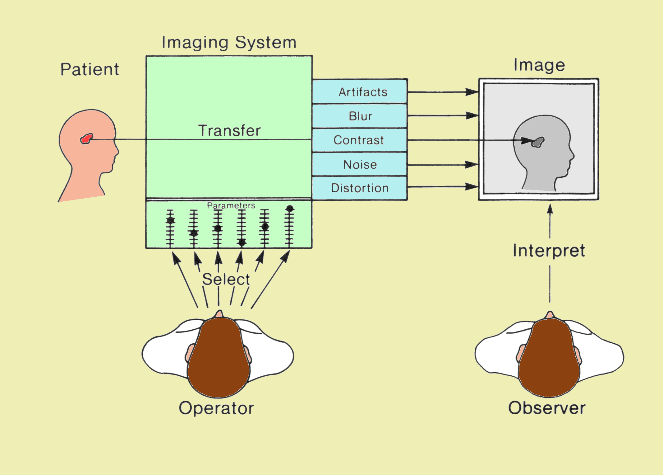
Components Associated with the Medical Imaging Process
The ability of an observer to detect signs of a pathologic process depends on a combination of three major factors: (1) image quality, (2) viewing conditions, and (3) observer performance characteristics.
|
|
|
|
|
CONTENTS |
|
The quality of a medical image is determined by the imaging method, the characteristics of the equipment, and the imaging variables selected by the operator. Image quality is not a single factor but is a composite of at least five factors: contrast, blur, noise, artifacts, and distortion, as shown
above. The relationships between image quality factors and imaging system variables are discussed in detail in later chapters.
The human body contains many structures and objects that are simultaneously imaged by most imaging methods. We often consider a single object in relation to its immediate background. In fact, with most imaging procedures the visibility of an object is determined by this relationship rather than by the overall characteristics of the total image.
Consider the figure below. The task of every imaging system is to translate a specific tissue characteristic into image shades of gray or color. If contrast is adequate, the object will be visible. The degree of contrast in the image depends on characteristics of both the object and the imaging system.
|
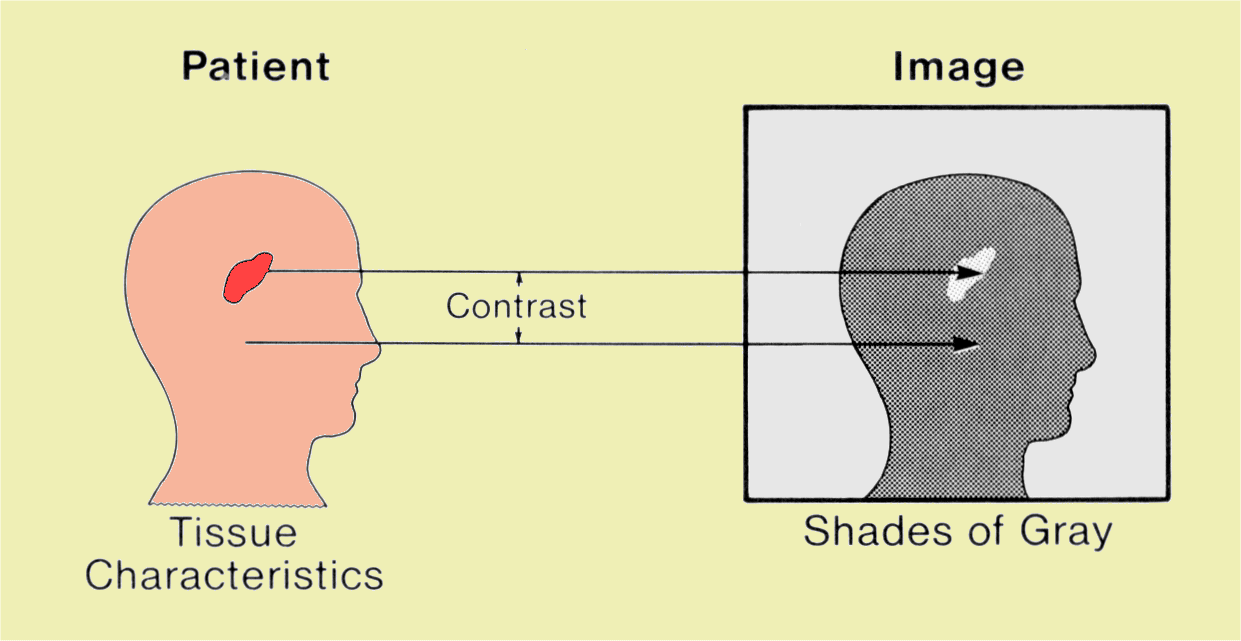
Medical Imaging is the Process of Converting Tissue Characteristics into a Visual Image
|
|
|
CONTENTS |
|
Contrast means difference. In an image, contrast can be in the form of different shades of gray, light intensities, or colors. Contrast is the most fundamental characteristic of an image. An object within the body will be visible in an image only if it has sufficient physical contrast relative to surrounding tissue. However, image contrast much beyond that required for good object visibility generally serves no useful purpose and in many cases is undesirable.
The physical contrast of an object must represent a difference in one or more tissue characteristics. For example, in radiography, objects can be imaged relative to their surrounding tissue if there is an adequate difference in either density or atomic number and if the object is sufficiently thick.
When a value is assigned to contrast, it refers to the difference between two specific points or areas in an image. In most cases we are interested in the contrast between a specific structure or object in the image and the area around it or its background.
|
|
|
|
|
CONTENTS |
|
The degree of physical object contrast required for an object to be visible in an image depends on the imaging method and the characteristics of the imaging system. The primary characteristic of an imaging system that establishes the relationship between image contrast and object contrast is its contrast sensitivity. Consider the situation shown
below. The circular objects are the same size but are filled with different concentrations of iodine contrast medium. That is, they have different levels of object contrast. When the imaging system has a relatively low contrast sensitivity, only objects with a high concentration of iodine (ie, high object contrast) will be visible in the image. If the imaging system has a high contrast sensitivity, the lower-contrast objects will also be visible.
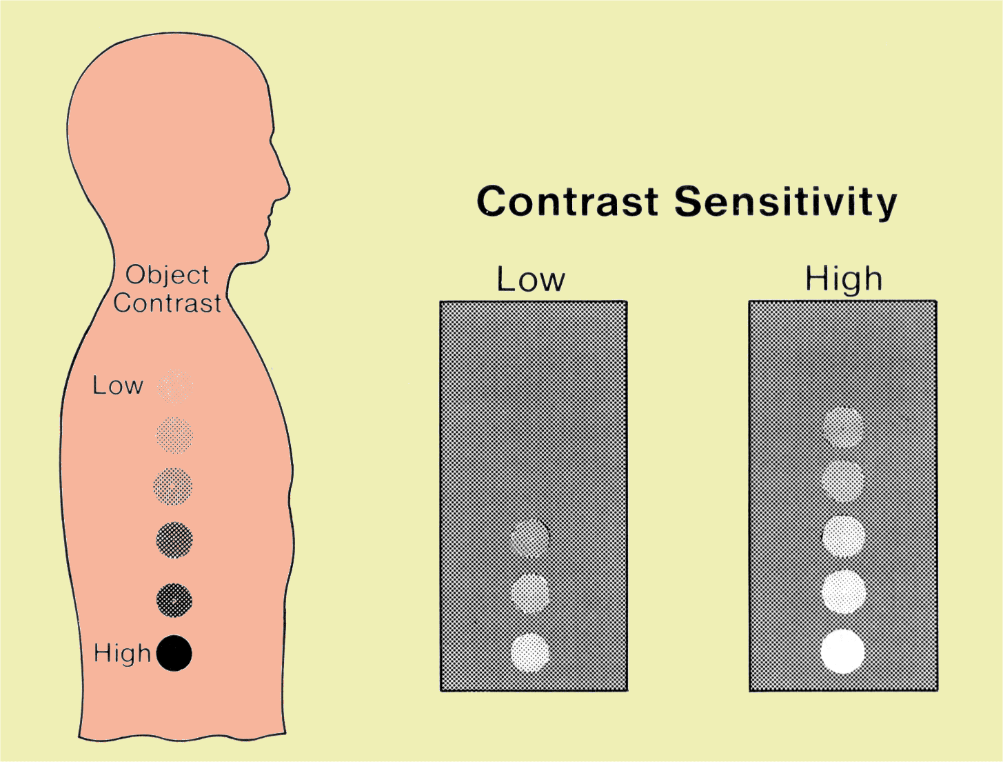
Increasing Contrast Sensitivity Increases Image Contrast and the Visibility of Objects in the Body
We emphasize that contrast sensitivity is a characteristic of the imaging method and the variables of the particular imaging system. It is the characteristic that relates to the system's ability to translate physical object contrast into image contrast. The contrast transfer characteristic of an imaging system can be considered from two perspectives. From the perspective of adequate image contrast for object visibility, an increase in system contrast sensitivity causes lower-contrast objects to become visible. However, if we consider an object with a fixed degree of physical contrast
(i.e., a fixed concentration of contrast medium), then increasing contrast sensitivity will increase image contrast.
It is difficult to compare the contrast sensitivity of various imaging methods because many are based on different tissue characteristics. However, certain methods do have higher contrast sensitivity than others. For example, computed tomography (CT) generally has a higher contrast sensitivity than conventional radiography. This is demonstrated by the ability of CT to image soft tissue objects (masses) that cannot be imaged with radiography. The specific factors that determine the contrast sensitivity of each imaging method are considered in later chapters.
Consider the image below. Here is a series of objects with different degrees of physical contrast. They could be vessels filled with different concentrations of contrast medium. The highest concentration (and contrast) is at the bottom. Now imagine a curtain coming down from the top and covering some of the objects so that they are no longer visible. Contrast sensitivity is the characteristic of the imaging system that raises and lowers the curtain. Increasing sensitivity raises the curtain and allows us to see more objects in the body. A system with low contrast sensitivity allows us to visualize only objects with relatively high inherent physical contrast.
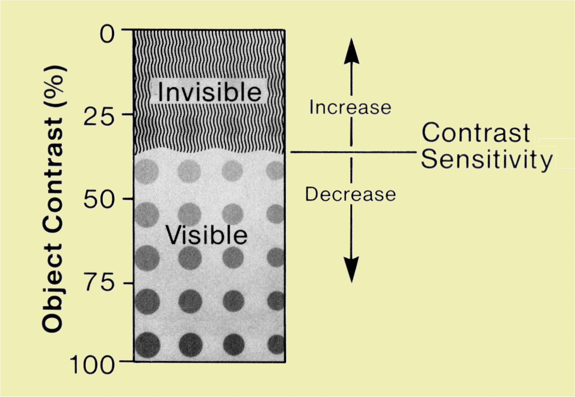
Effect of Contrast Sensitivity on Object Visibility
|
|
|
|
|
CONTENTS |
|
Structures and objects in the body vary not only in physical contrast but also in size. Objects range from large organs and bones to small structural features such as trabecula patterns and small calcifications. It is the small anatomical features that add detail to a medical image. Each imaging method has a limit as to the smallest object that can be imaged and thus on visibility of detail. Visibility of detail is limited because all imaging methods introduce blurring into the process. The primary effect of image blur is to reduce the contrast and visibility of small objects or detail.
Consider the image below, which represents the various objects in the body in terms of both physical contrast and size. As we said, the boundary between visible and invisible objects is determined by the contrast sensitivity of the imaging system. We now extend the idea of our curtain to include the effect of blur. It has little effect on the visibility of large objects but it reduces the contrast and visibility of small objects. When blur is present, and it always is, our curtain of invisibility covers small objects and image detail. Blur and visibility of detail are discussed in more depth in
a later chapter.
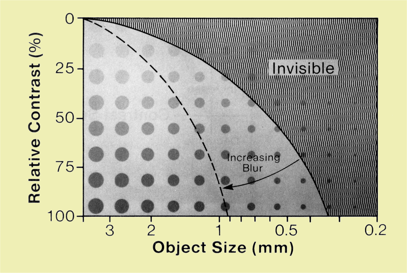
Effect of Blur on Visibility of Image Detail
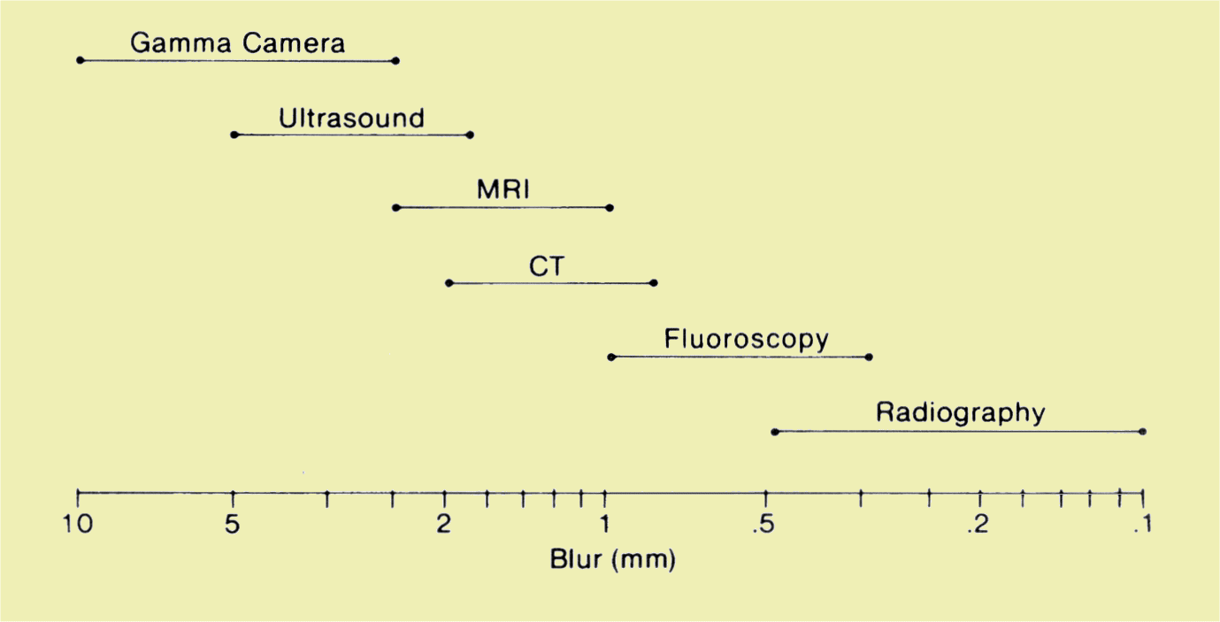
The amount of blur in an image can be quantified in units of length. This value represents the width of the blurred image of a small object.
The image below compares the approximate blur values for medical imaging methods. As a general rule, the smallest object or detail that can be imaged has approximately the same dimensions as those of the image blur.
|
Range of Blur Values and Visibility of Detail Obtained with Various Imaging Methods
|
|
|
CONTENTS |
|
Another characteristic of all medical images is image noise. Image noise, sometimes referred to as image mottle, gives an image a textured or grainy appearance. The source and amount of image noise depend on the imaging method and are discussed in more detail in
a later chapter. We now briefly consider the effect of image noise on visibility.
In the image below we find our familiar array of body objects arranged according to physical contrast and size. We now add a third factor, noise, which will affect the boundary between visible and invisible objects. The general effect of increasing image noise is to lower the curtain and reduce object visibility. In most medical imaging situations the effect of noise is most significant on the low-contrast objects that are already close to the visibility threshold.
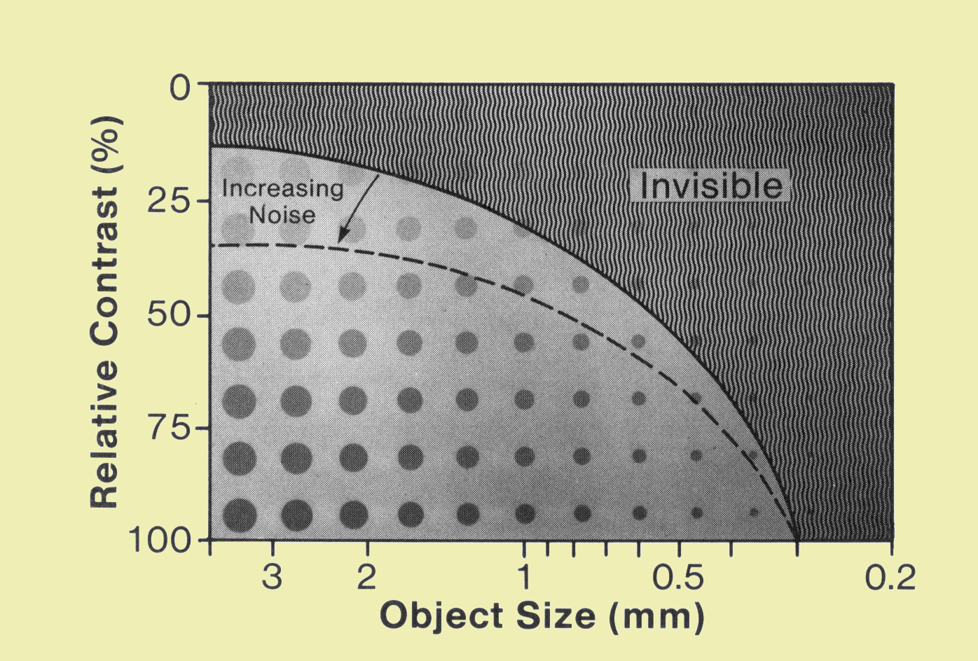
|
Effect of Noise on Object Visibility
|
|
|
CONTENTS |
|
We have seen that several characteristics of an imaging method (contrast sensitivity, blur, and noise) cause certain body objects to be invisible. Another problem is that most imaging methods can create image features that do not represent a body structure or object. These are image artifacts. In many situations an artifact does not significantly affect object visibility and diagnostic accuracy. But artifacts can obscure a part of an image or may be interpreted as an anatomical feature. A variety of factors associated with each imaging method can cause image artifacts.
|
|
|
|
|
CONTENTS |
|
A medical image should not only make internal body objects visible, but should give an accurate impression of their size, shape, and relative positions. An imaging procedure can, however, introduce distortion of these three factors.
|
|
|
|
|
CONTENTS |
|
It would be logical to raise the question as to why we do not adjust each imaging procedure to yield maximum visibility. The reason is that in many cases the variables that affect image quality also affect factors such as radiation exposure to the patient and imaging time. In general, an imaging procedure should be set up to produce adequate image quality and visibility without excessive patient exposure or imaging time.
In many situations, if a variable is changed to improve one characteristic of image quality, such as noise, it often adversely affects another characteristic, such as blur and visibility of detail. Therefore an imaging procedure must be selected according to the specific requirements of the clinical examination.
|
|
|
|
|
CONTENTS |
|
A combination of two factors makes each imaging method unique. These are the tissue characteristics that are visible in the image and the viewing perspective. The specific tissue characteristics that produce the various shades of gray and image contrast vary among the various modalities and methods.
A radiologist uses an image to search for signs of a pathologic condition or injury in the body. Signs can be observed only if the condition produces a physical change in the associated tissue. Many pathologic conditions produce a change in a physical characteristic that can be imaged by one method but not another.
Imaging methods create images that show the body from one of two perspectives, through either projection or tomographic imaging. There are advantages and disadvantages to each.
In projection imaging (radiography and fluoroscopy), images are formed by projecting an x-ray beam through the patient's body and casting shadows onto an appropriate receptor that converts the invisible x-ray image into a visible light image. The gamma camera records a projection image that represents the distribution of radioactive material in the body. The primary advantage of this type of image is that a large volume of the patient's body can be viewed with one image. A disadvantage is that structures and objects are often superimposed so that the image of one might interfere with the visibility of another. Projection imaging produces spatial distortion that is generally not a major problem in most clinical applications.
Tomographic imaging, i.e., conventional tomography, computed tomography (CT), sonography, single photon emission tomography (SPECT), positron emission tomography (PET), and MRI, produces images of selected planes or slices of tissue in the patient's body. The general advantage of a tomographic image is the increased visibility of objects within the imaged plane. One factor that contributes to this is the absence of overlying objects. The major disadvantage is that only a small slice of a patient's body can be visualized with one image. Therefore, most tomographic procedures usually require many images to survey an entire organ system or body cavity.
|
|
|
|
|
CONTENTS |
|
Our ability to see a specific object or feature in an image depends on the conditions under which we view the image. We must deal with the effects of viewing conditions in many activities in addition to the professional interpretation of medical images. Dim candlelight enhances the pleasure of a fine dinner but often makes it difficult to read the menu. The glare of an oncoming automobile headlight reduces our ability to see objects in the road and also produces discomfort and stress. We quickly learn that there is an optimum viewing distance for television sets, newspapers, etc. A small object dropped onto the smooth surface of the dining table is easier to see than an object dropped onto a textured carpet or sandy beach. With these experiences in mind, let us consider the factors associated with image viewing conditions and how they affect our ability to visualize body structures.
The image below shows the primary factors that affect our ability to see or detect an object in an image. We will assume a circular object located within a larger background area. The ability of an observer to detect the object depends on a combination of factors including object contrast and size, background, brightness (luminance) and structure (texture), glare produced by other light sources, distance between the image and the observer, and the time available to search for the object.
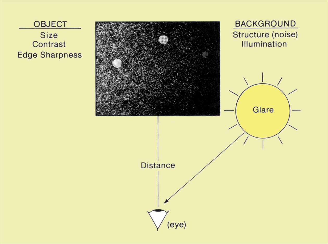
Viewing Condition Factors That Affect Object Visibility
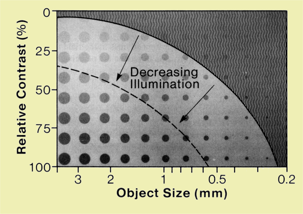
The image below is an image of the array of objects we used to demonstrate the effects of image quality factors. We now use it to demonstrate how the factors associated with the viewing process affect our ability to see the objects. You can use this actual image to test the factors discussed below.
|
Effect of Viewing Conditions on Object Visibility
|
|
|
CONTENTS |
|
The ability to see or detect an object is heavily influenced by the contrast between the object and its background. For most viewing tasks there is not a specific threshold contrast at which the object suddenly becomes visible. Instead, the accuracy of seeing or detecting a specific object increases with contrast.
The contrast sensitivity of the human viewer changes with viewing conditions. When viewer contrast sensitivity is low, an object must have a relatively high contrast to be visible. The degree of contrast required depends on conditions that alter the contrast sensitivity of the observer: background brightness, object size, viewing distance, glare, and background structure.
|
|
|
|
|
CONTENTS |
|
The human eye can function over a large range of light levels or brightness, but vision is not equally sensitive at all brightness levels. The ability to detect objects generally increases with increasing background brightness or image illumination. To be detected in areas of low brightness, an object must be large and have a relatively high level of contrast with respect to its background. This can be demonstrated with the image in
the image above. View this image with different levels of illumination. You will notice that under low illumination you cannot see all of the small and low-contrast objects. A higher level of object contrast is required for visibility.
Viewbox luminance (brightness) can have a significant effect on the visibility of objects within an image. For general image viewing, viewboxes should have a luminance of at least 1,500 nits. A brighter viewbox of at least 3,500 nits is recommended for mammography. A nit is a unit of brightness and is described in more detail in
a later chapter.
|
|
|
|
|
CONTENTS |
|
The relationship between the degree of contrast required for detectability and for background brightness is influenced by the size of the object. Small objects require either a higher level of contrast or increased background brightness to be detected.
The detectability of an object is more closely related to the angle it forms in the visual field. The angle is the ratio of object diameter to the distance between image and observer. In principle, a small object will have the same detectability at close range as a larger object viewed at a greater distance.
|
|
|
|
|
CONTENTS |
|
The relationship between visibility and viewing distance is affected by several factors. When the viewing distance is reduced, an object creates a larger angle and is generally easier to see. However, the eye does not focus and exhibit maximum contrast sensitivity at close range. Therefore, the relationship between detectability and viewing distance generally peaks at a distance of approximately 2 ft.
|
|
|
CONTENTS |
|
Glare is produced by bright areas or light sources in the field of view and has several undesirable effects. One effect is to reduce the perceived contrast of the objects viewed. When light from the glare-producing source enters the eye, some of it is scattered over other areas within the visual field. This in turn reduces contrast sensitivity. The extent to which it is reduced depends on the brightness and size of the glare source and its proximity to the object viewed.
Glare is a major problem when some of the bright viewbox area is not covered by the film. Visibility is improved by masking when viewing small films as in mammography.
|
|
|
CONTENTS |
|
The structure or texture of an object's background has a significant effect on its visibility. A smooth background produces maximum visibility; visibility of low contrast objects is often reduced because of the texture of surrounding tissues or image noise.
|
|
|
CONTENTS |
|
In many situations, the presence of a specific object or sign is not obvious but requires establishment by a trained observer. The criteria used to establish the presence of a specific sign often vary among observers. Individual observers also use different criteria, often influenced by the clinical significance of a specific observation.
|
|
|
CONTENTS |
|
Let us assume we have a relatively large number of cases to be examined by
means of a medical imaging procedure, and that a specific pathologic
condition is present in some and absent in others. The ideal situation
would be if the condition were diagnosed as positive when present and
negative when absent. In actual practice, this is usually not achieved. A
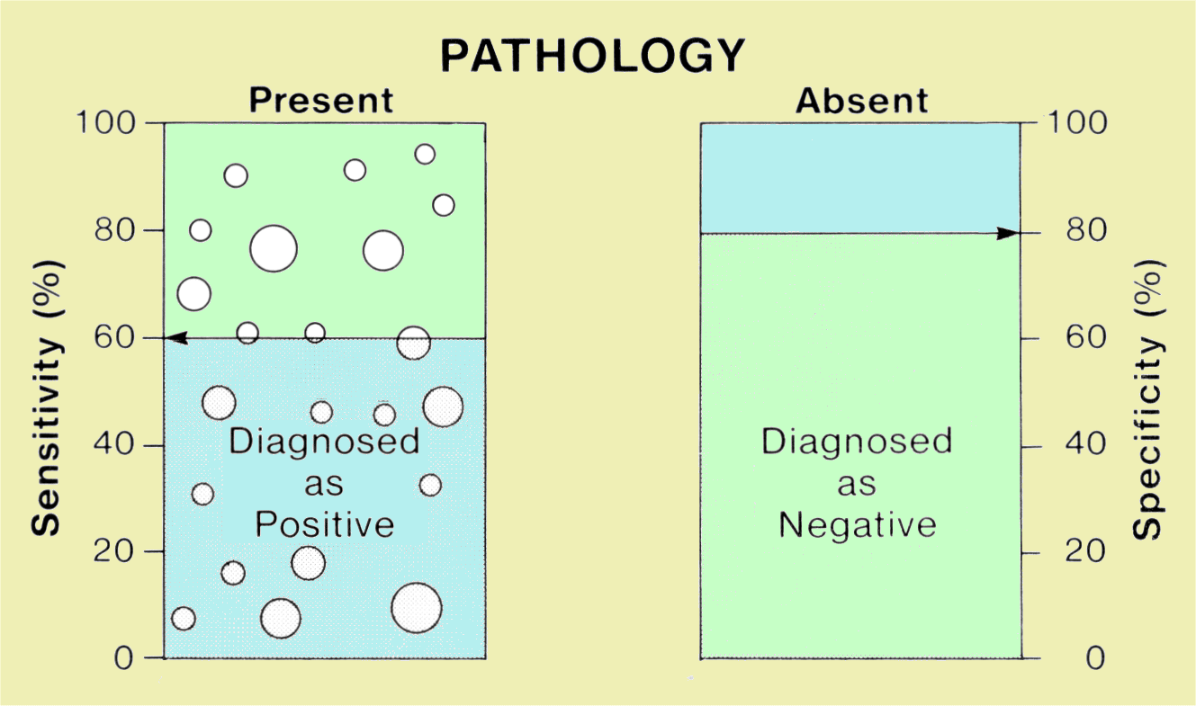
more realistic situation is represented in the figure below. Here we see
that a fraction of the pathological conditions were diagnosed as positive.
This fraction (or percentage) represents the sensitivity of the specific
diagnostic procedure. We also see that the condition was not always
diagnosed as negative when absent. The percentage of these cases diagnosed
as negative represents the specificity of the procedure.
Sensitivity and Specificity
|
|
|
CONTENTS |
|
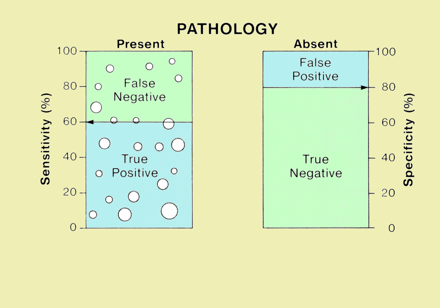
The diagnoses derived from the imaging procedure divide the cases
into four categories, as shown in the image below: true positives, true
negatives, false positives, and false negatives. In the ideal situation,
there are only true positives and true negatives. This would be a
diagnostic process with 100% accuracy.
Relationship of True and False Diagnostic Decisions to Sensitivity and Specificity
False negatives and false positives occur for a number of reasons,
including inherent limitations of a specific imaging method, selection of
inappropriate imaging factors, poor viewing conditions, and the
performance of the observer (radiologist). |
|
|
CONTENTS |
|
In general, if an observer is aggressive in trying to increase the
number of true positives (sensitivity), the number of false negatives
(decreased specificity) also increases. The relationship between
sensitivity and specificity for a specific diagnostic test (including
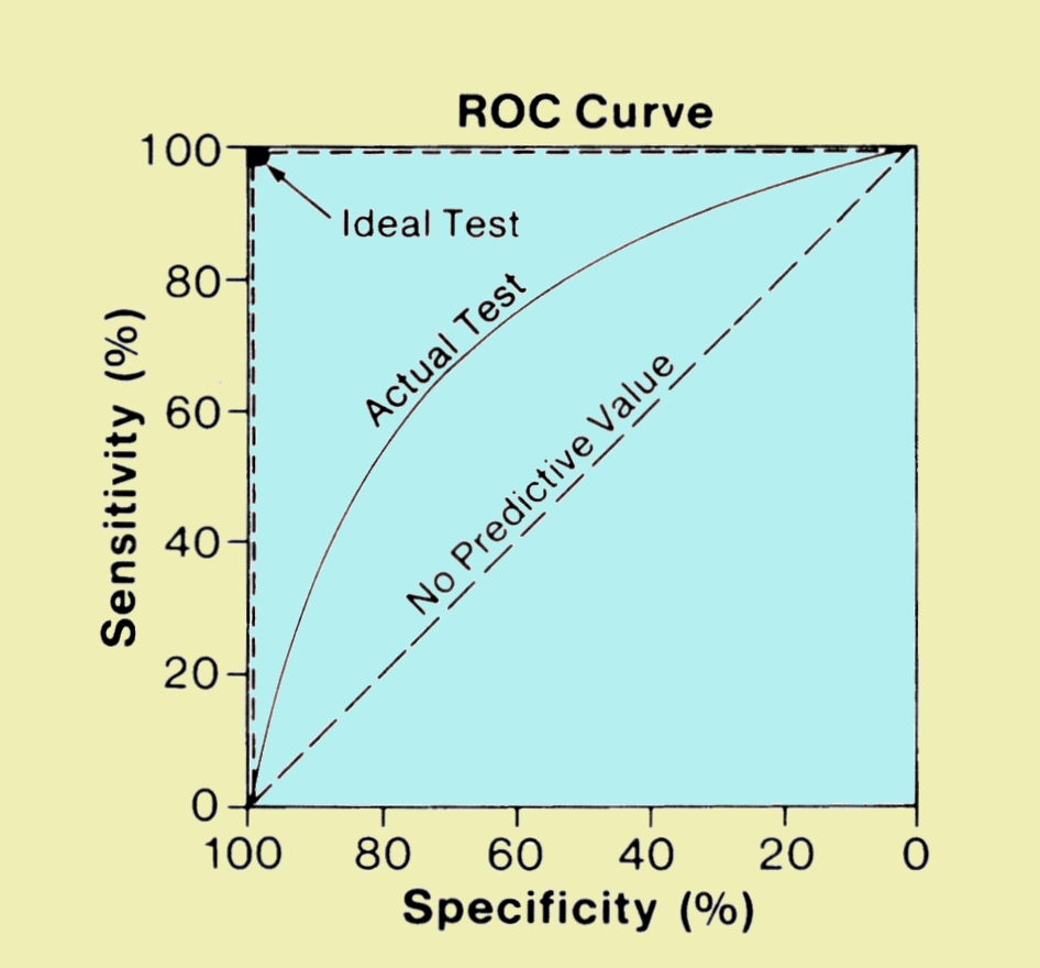
observer performance) can be described by a graph (shown in the figure
below) known as a receiver operating characteristic (ROC) curve.
Comparison of ROC Curves for an Ideal Diagnostic Procedure with One that Produces No Useful Information
The ideal diagnostic test produces 100% sensitivity and 100% specificity as shown. If a diagnostic procedure has no predictive value, and the diagnosis is obtained by a random selection process, the relationship between sensitivity and specificity is linear as shown. The observer determines the actual operating point along this line. Since this particular diagnostic procedure is providing no useful information, an attempt to increase the sensitivity by calling a greater number of positives will produce a proportionate decrease in the specificity.
|
|
|
CONTENTS |
|
The relationship between sensitivity and specificity for most
medical imaging procedures is between the ideal and no predictive value.
The ROC curve shown below is typical. The characteristics of the imaging
method and the quality of the resulting image determine the shape of the
curve and the relationship between sensitivity and specificity for a
specific pathological condition. The criteria used by the observer to make
the diagnosis determine the point on the curve that produces the actual
sensitivity and specificity values.
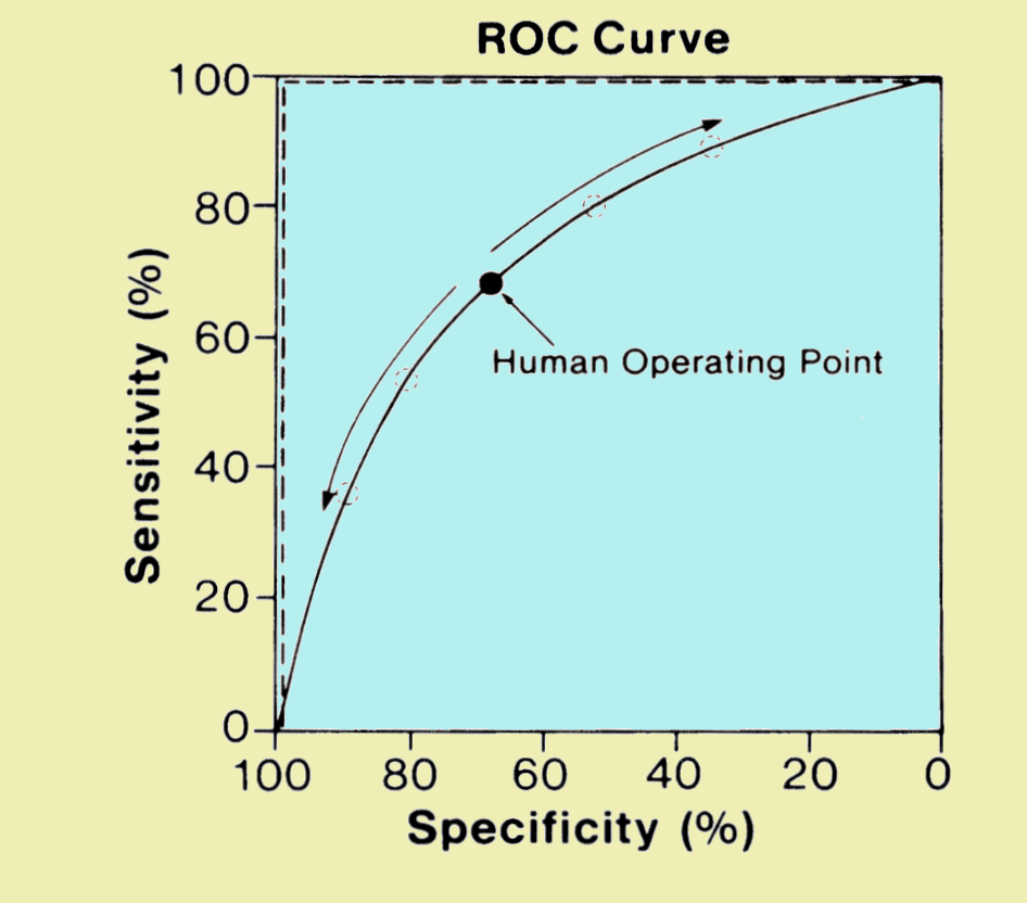
An ROC Curve for a Specific Imaging Procedure. The Actual Operating Point is Determined by Characteristics of the Observer.
|
|