
Chapter 8
|
|
Chapter 8 |
| Link to Book Table of Contents | Chapter Contents Shown Below |
Fat and fluid are two materials in the body that can produce very intense
signals and brightness in images. This occurs with fat in T1 images and with
fluid in T2 images. A possible problem is that these bright regions can reduce
the visibility of other tissues and pathologic conditions in the area.
T1-Based Fat And Fluid Suppression
|
Figure 8-1. The use
of STIR to suppress signals from fat by setting TI to a value (short)
that will image the longitudinal magnetization at the time when fat is
relaxing through the zero level. |
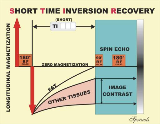 |
The
TI interval is selected so that the “picture is snapped” by applying the
excitation pulse at that time. Because the fat has no magnetization at that
time, it will not produce a signal. Since this is achieved with relatively short
values for TI, this method of fat suppression is often referred to as Short Time
Inversion Recovery (STIR).
STIR is just the inversion recovery (IR) method with the TI set to a
relatively low value. The description of the basic IR method in Chapter 6 shows
how the factor TI is used to select the time at which the longitudinal
magnetization “picture is snapped” and the magnetization is converted into image
contrast. The ability to use this method to suppress the signals from fat is
based on the fact that the longitudinal magnetization of fat passes through zero
at a time before and separated from the other tissues. Setting the TI to measure
the longitudinal magnetization at the time when fat is at zero produces no
signal and fat will be dark in the image.
The best TI value to suppress the signals from fat depends on the T1
value of fat, which depends on the strength of the magnetic field. It will
generally be in the range of 120 to 150 msec for field strengths in the 0.5 T to
1.5 T range.
Another consideration with STIR is that the TR must be set relatively
long (1500–2000 msec), compared to a T1 image acquisition with spin echo using a
TR value of approximately 500 msec. This additional time is required for the
longitudinal magnetization to more fully recover after the excitation pulse and
before the next cycle can begin.
|
Figure 8-2. The suppression of fluid by
selecting a long TI that will image the longitudinal magnetization at
the time when fluid is relaxing through zero. |
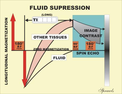 |
This
works because the long T1 values of fluids are well separated from the T1 values
of other tissues. By setting the TI to a long value as shown, the longitudinal
magnetization is converted to transverse and the “picture is snapped” when the
fluid is at a zero value. Fluids appear as dark regions in the image. When fluid
suppression is used with a T2 image acquisition (long TE), the usually bright
fluid is suppressed but other tissues with long T2 values, such as pathologic
tissue, remain bright.
Acquisition time is a special concern with this method. That is because
when long TI values are used, the TR values must also be long (5000–6000 msec)
and that increases the acquisition time. For this reason, the practical thing is
to use this method with one of the fast acquisition techniques.
|
Figure 8-3.
Suppressing the signals from fat by applying an inversion pulse tuned to
the resonant frequency of fat so that it does not affect the other
tissues. |
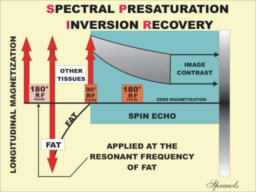 |
The
unique feature of this method is that the imaging cycle begins with an inversion
pulse that is applied at the fat resonant frequency. This selectively inverts
the longitudinal magnetization of the fat without affecting the other tissues.
The TI is set so that the spin echo excitation pulse is applied at the time when
the fat longitudinal magnetization is passing through zero. This results in T1
and T2 images with the signals from fat removed.
The advantage of the SPIR method is that the contrast of tissues with
relatively short T1 values is not diminished as it might be with the STIR
method. For example, the use of gadolinium contrast media reduces the T1 value
of the water component of tissue. These short T1 value signals would be
suppressed by STIR, but not by SPIR.
There are some precautions that must be observed when using SPIR. They
relate to having very good magnetic field homogeneity. Recall that the resonant
frequency is controlled by the field strength in each location. Therefore, for
the RF suppression pulse to accurately suppress the fat magnetization over the
image area, the fat must be resonating at precisely the same frequency. This
requires a very homogeneous (within just a few parts per million) magnetic
field. This is achieved by shimming the field before the acquisition, removing
metal objects that might distort the field, and by using a relative small field
of view.
An alternative to the SPIR method is to apply a saturation rather than an
inversion pulse tuned to the fat resonant frequency. This is sometimes referred
to as chemical saturation.
Magnetization Transfer Contrast
(MTC)
Magnetization Transfer Contrast (MTC) is a technique that enhances image
contrast by selectively suppressing the signals from specific tissues. The
amount of suppression depends on a specific tissue’s magnetization transfer
characteristics. Maximum suppression is obtained for tissues that have a
high level of magnetization transfer.
The MTC technique is illustrated in Figure 8-4.
|
Figure 8-4. The use of magnetization
transfer between different types of tissue to suppress selective
signals. |
 |
It
is based on the principle that the protons in tissue are in different states of
mobility, which we will designate as the “free” pool and the “bound” pool.
The
protons that produce signals and are visible in MRI are not rigidly bound and
might be considered to be “free” and in a general “semi-solid” structure. This
environment produces relatively long T2 values (in comparison to the bound
state) and a relatively narrow resonant frequency.
Most
tissues also contain protons that are more rigidly bound and associated with
more “solid” structures such as large macromolecules and membranes. These
structures have very short T2 values. This means that the transverse
magnetization decays before it can be imaged with the usual methods. Therefore,
these protons do not contribute to the image. An important characteristic of
these protons is that they have a much broader resonant frequency spectrum than
the “free” protons.
Prior to the beginning of the imaging acquisition cycle a saturation
pulse is applied at a frequency that is different from the resonant frequency of
the “free” protons. Therefore, it does not have a direct effect on the protons
that are producing the signals. However, the saturation pulse is within the
broader resonant frequency of the “bound” protons. It produces saturation of the
longitudinal magnetization in the “bound” pool.
The effect of the saturation is now transferred to the longitudinal
magnetization of the “free” pool by the magnetization transfer process. The key
is that the transfer is not the same for all tissues. Only the tissues with a
relatively high magnetization transfer coupling and a significant bound pool
concentration will experience the saturation and have their signals reduced in
intensity.
Fluids, fat, and bone marrow have very little, if any, magnetization
transfer. Therefore, they will not experience the transferred saturation, and
will remain relatively bright in the images.
Most other tissues have some, but varying degrees of, magnetization
transfer. When the MTC technique is used, the saturation produced by the RF
pulse applied to the “bound” protons will be transferred to the “free” protons,
but only in those tissues that have a significant magnetization transfer
capability. The result is that these tissues will be saturated to some degree
and their signal intensities will be reduced.
Therefore, MTC is a way of enhancing contrast in an image by suppressing
the signals from tissues that have a relatively high magnetization transfer. One
example is to use MTC to reduce the brightness (signal intensity) of brain
tissue so that the vascular structures will be brighter in angiography.
There
are procedures in which it is desirable to suppress signals from specific
anatomical regions. The two major applications of this are to reduce
motion-induced artifacts, as described in Chapter 14, and to suppress the
signals from blood that is flowing in a specific direction, as discussed in
Chapter 12. At this time we will consider the general technique, which is
illustrated in Figure 8-5.
|
Figure 8-5. The
application of a saturation pulse can be directed to a specific
anatomical region to suppress undesirable signals from moving tissues. |
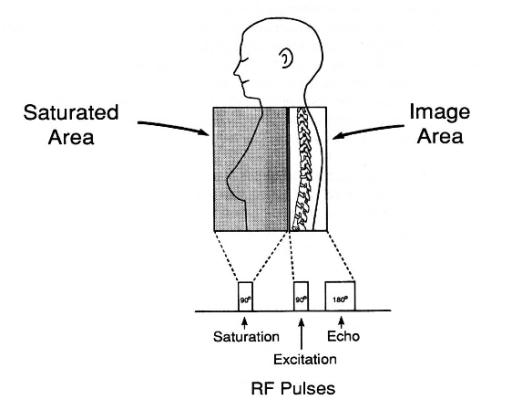 |
Let us recall that gradients are used to vary the magnetic field strength
across a patient’s body. In the
presence of a gradient one region of the body is in a different field strength
from another and is therefore tuned to a different resonant frequency. This
makes it possible to apply RF pulses selectively to specific regions without
affecting adjacent regions.
In Chapter 14 we will see that a major source of artifacts in MRI is the
motion or movement of tissues and fluids. The motion produces errors in the
spatial encoding of the signals that causes them to be displayed in the wrong
location in the image. Signals from moving tissues and fluids are displayed as
streaks, which are undesirable artifacts.
With the regional saturation technique the objective is to suppress
selective signals originating from one region, usually the moving tissue or
fluid, without affecting these signals in the region that is being imaged. The
specific applications of this will be described in Chapter 14.
Prior to the imaging cycle pulse sequence, a saturation pulse is
selectively applied to the region that is to be suppressed. The saturation pulse
is given a frequency that is different from the frequency of the other imaging
pulses. This is so that it will be tuned to the resonant frequency of the region
that is to be suppressed. This region will have a resonant frequency different
from the imaged area because of the presence of the gradient as described above.
The region that is saturated is a three-dimensional (3-D) volume or slab
of tissue. It is important that the slab be properly positioned in relationship
to the imaged area for best results.
The
application of regional saturation to suppress artifacts will be discussed in
more detail when we consider artifacts in Chapter 14.
Mind Map Summary
Selective Signal Suppression
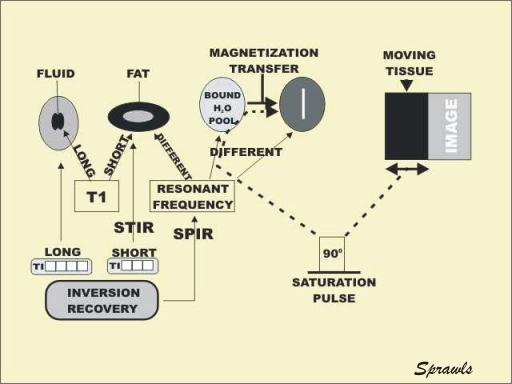
It is often desirable to suppress the signals and resulting brightness of
selected tissues or anatomical regions to improve visibility of other tissues or
general image quality. It is possible to selectively suppress signals from
specific tissues if the tissues are significantly different from the other
tissues in terms of some MR characteristic.
Signals from fat, generally very bright in T1 images, can be suppressed
with two techniques. Because fat has a very short T1 value compared to other
tissues, it can be suppressed with the STIR method, an inversion recovery method
in which the TI is set to snap the picture when the magnetization of fat is
passing through the zero level. The resonant frequency of fat molecules is
slightly different from water molecules because of the chemical shift effect.
The SPIR method makes use of this by applying an RF pulse at the fat frequency
to reduce the fat magnetization to the zero level at the beginning of each
imaging cycle.
Signals from fluid can be suppressed by using an inversion recovery
method with the TI set to a long value. This works because fluids have long T1
values and the fluid’s magnetization passes through the zero level significantly
later and separate from that of tissues. The MTC technique can be used to reduce
signal intensity from tissues that have a relatively high magnetization transfer
characteristic. This can be used to enhance image contrast.
Saturation pulses can be selectively applied to specific anatomical
regions to suppress any signals that could occur from tissues or fluids in that
region. This is useful for reducing motion artifacts and also for reducing the
signals from flowing blood in specific anatomical regions.